5J5E

crystal structure of antigen-ERAP1 domain complex


Experimental Data Snapshot

  • Method: X-RAY DIFFRACTION
  • Resolution: 2.80 Å
  • R-Value Free: 0.269 
  • R-Value Work: 0.207 
  • R-Value Observed: 0.210 

wwPDB Validation   3D Report Full Report


This is version 1.5 of the entry. See complete history


Literature

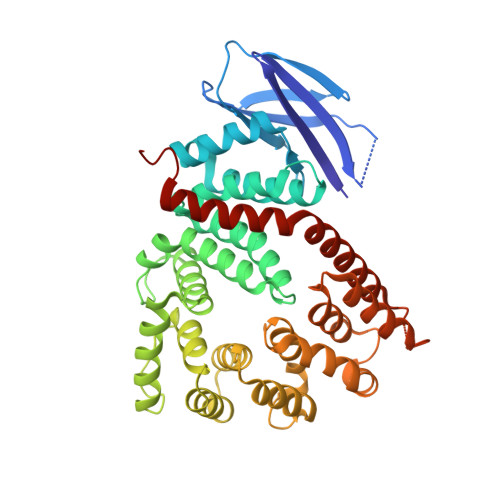
Crystal structure of a polypeptide's C-terminus in complex with the regulatory domain of ER aminopeptidase 1.

Sui, L.Gandhi, A.Guo, H.C.

(2016) Mol Immunol 80: 41-49

  • DOI: https://doi.org/10.1016/j.molimm.2016.10.012
  • Primary Citation of Related Structures:  
    5J5E

  • PubMed Abstract: 

    Endoplasmic reticulum aminopeptidase 1 (ERAP1) is involved in the final processing of peptide precursors to generate the N-termini of MHC class I-restricted epitopes. ERAP1 thus influences immunodominance and cytotoxic immune responses by controlling the peptide repertoire available for cell surface presentation by MHC molecules. To enable this critical role in antigen processing, ERAP1 trims peptides by a unique molecular ruler mechanism that turns on/off hydrolysis activity in a peptide-length and -sequence dependent manner. Thus unlike other aminopeptidases, ERAP1 could recognize both the N- and C-termini of peptides in order to read the substrate's length. To exemplify and validate this molecular ruler mechanism, we have carried out crystallographic studies on molecular recognition of antigenic peptide's C-terminus by ERAP1. In this report, we have determined a 2.8Å-resolution crystal structure of an intermolecular complex between the ERAP1 regulatory domain and a natural epitope's C-terminus displayed in a fusion protein. It reveals the structural details of peptide's C-termini recognition by ERAP1. ERAP1 uses specificity pockets on the regulatory domain to bind the peptide's carboxyl end and side chain of the C-terminal anchoring residue. At the same time, flexibility in length and sequence at the middle of peptides is accommodated by a kink with minimal interactions with ERAP1.


  • Organizational Affiliation

    Department of Biological Sciences, University of Massachusetts Lowell, 1 University Avenue, Lowell, MA 01854, USA.


Macromolecules
Find similar proteins by:  (by identity cutoff)  |  3D Structure
Entity ID: 1
MoleculeChains Sequence LengthOrganismDetailsImage
Endoplasmic reticulum aminopeptidase 1420Homo sapiensMutation(s): 0 
Gene Names: ERAP1APPILSARTS1KIAA0525UNQ584/PRO1154
EC: 3.4.11
UniProt & NIH Common Fund Data Resources
Find proteins for Q9NZ08 (Homo sapiens)
Explore Q9NZ08 
Go to UniProtKB:  Q9NZ08
PHAROS:  Q9NZ08
GTEx:  ENSG00000164307 
Entity Groups  
Sequence Clusters30% Identity50% Identity70% Identity90% Identity95% Identity100% Identity
UniProt GroupQ9NZ08
Sequence Annotations
Expand
  • Reference Sequence
Experimental Data & Validation

Experimental Data

  • Method: X-RAY DIFFRACTION
  • Resolution: 2.80 Å
  • R-Value Free: 0.269 
  • R-Value Work: 0.207 
  • R-Value Observed: 0.210 
  • Space Group: P 1 21 1
Unit Cell:
Length ( Å )Angle ( ˚ )
a = 64.26α = 90
b = 66.85β = 110.16
c = 66.5γ = 90
Software Package:
Software NamePurpose
REFMACrefinement
iMOSFLMdata reduction
xia2data reduction
PHASERphasing

Structure Validation

View Full Validation Report


Entry History & Funding Information

Deposition Data


Funding OrganizationLocationGrant Number
National Institutes of Health/National Institute Of Allergy and Infectious Diseases (NIH/NIAID)United StatesAI068831
National Institutes of Health/National Institute Of Allergy and Infectious Diseases (NIH/NIAID)United StatesAI078134

Revision History  (Full details and data files)

  • Version 1.0: 2017-04-12
    Type: Initial release
  • Version 1.1: 2017-09-20
    Changes: Advisory, Author supporting evidence, Refinement description
  • Version 1.2: 2019-12-11
    Changes: Author supporting evidence
  • Version 1.3: 2021-05-19
    Changes: Database references
  • Version 1.4: 2022-04-27
    Changes: Advisory, Database references
  • Version 1.5: 2023-09-27
    Changes: Data collection, Refinement description