5FV9

Crystal structure of GalNAc-T2 in complex with compound 16d


Experimental Data Snapshot

  • Method: X-RAY DIFFRACTION
  • Resolution: 2.07 Å
  • R-Value Free: 0.247 
  • R-Value Work: 0.190 
  • R-Value Observed: 0.191 

wwPDB Validation   3D Report Full Report


Ligand Structure Quality Assessment 


This is version 1.2 of the entry. See complete history


Literature

Glycomimetics Targeting Glycosyltransferases: Synthetic, Computational and Structural Studies of Less-Polar Conjugates.

Ghirardello, M.De Las Rivas, M.Lacetera, A.Delso, I.Lira-Navarrete, E.Tejero, T.Martin-Santamaria, S.Hurtado-Guerrero, R.Merino, P.

(2016) Chemistry 22: 7215

  • DOI: https://doi.org/10.1002/chem.201600467
  • Primary Citation of Related Structures:  
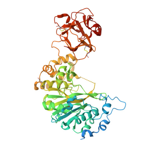
    5FV9

  • PubMed Abstract: 

    The Leloir donors are nucleotide sugars essential for a variety of glycosyltransferases (GTs) involved in the transfer of a carbohydrate to an acceptor substrate, typically a protein or an oligosaccharide. A series of less-polar nucleotide sugar analogues derived from uridine have been prepared by replacing one phosphate unit with an alkyl chain. The methodology is based on the radical hydrophosphonylation of alkenes, which allows coupling of allyl glycosyl compounds with a phosphate unit suitable for conjugation to uridine. Two of these compounds, the GalNAc and galactose derivatives, were further tested on a model GT, such as GalNAc-T2 (an important GT widely distributed in human tissues), to probe that both compounds bound in the medium-high micromolar range. The crystal structure of GalNAc-T2 with the galactose derivative traps the enzyme in an inactive form; this suggests that compounds only containing the β-phosphate could be efficient ligands for the enzyme. Computational studies with GalNAc-T2 corroborate these findings and provide further insights into the mechanism of the catalytic cycle of this family of enzymes.


  • Organizational Affiliation

    Departamento de Síntesis y Estructura de Biomoléculas, Instituto de Síntesis Química y Catálisis Homogénea (ISQCH), Universidad de Zaragoza, CSIC, 50009, Zaragoza, Aragón, Spain.


Macromolecules
Find similar proteins by:  (by identity cutoff)  |  3D Structure
Entity ID: 1
MoleculeChains Sequence LengthOrganismDetailsImage
GALNAC-T2
A, B, C, D, E
A, B, C, D, E, F
571Homo sapiensMutation(s): 1 
UniProt & NIH Common Fund Data Resources
Find proteins for Q10471 (Homo sapiens)
Explore Q10471 
Go to UniProtKB:  Q10471
PHAROS:  Q10471
GTEx:  ENSG00000143641 
Entity Groups  
Sequence Clusters30% Identity50% Identity70% Identity90% Identity95% Identity100% Identity
UniProt GroupQ10471
Sequence Annotations
Expand
  • Reference Sequence
Small Molecules
Ligands 5 Unique
IDChains Name / Formula / InChI Key2D Diagram3D Interactions
Y6W
Query on Y6W

Download Ideal Coordinates CCD File 
BA [auth E],
IA [auth F],
N [auth B]
[(2R,3S,4R,5R)-5-(2,4-dioxo-3,4-dihydropyrimidin-1(2H)-yl)-3,4-dihydroxytetrahydrofuran-2-yl]methyl hydrogen (S)-{3-[(2R,3R,4R,5R,6R)-3,4,5-trihydroxy-6-(hydroxymethyl)tetrahydro-2H-pyran-2-yl]propyl}phosphonate
C18 H29 N2 O13 P
PDUJCKZVDKZKPQ-RUIRHDHUSA-N
UDP
Query on UDP

Download Ideal Coordinates CCD File 
G [auth A]
GA [auth F]
P [auth B]
S [auth C]
X [auth D]
G [auth A],
GA [auth F],
P [auth B],
S [auth C],
X [auth D],
Z [auth E]
URIDINE-5'-DIPHOSPHATE
C9 H14 N2 O12 P2
XCCTYIAWTASOJW-XVFCMESISA-N
GOL
Query on GOL

Download Ideal Coordinates CCD File 
L [auth A],
M [auth A],
MA [auth F],
NA [auth F],
W [auth C]
GLYCEROL
C3 H8 O3
PEDCQBHIVMGVHV-UHFFFAOYSA-N
EDO
Query on EDO

Download Ideal Coordinates CCD File 
CA [auth E]
DA [auth E]
EA [auth E]
FA [auth E]
I [auth A]
CA [auth E],
DA [auth E],
EA [auth E],
FA [auth E],
I [auth A],
J [auth A],
JA [auth F],
K [auth A],
KA [auth F],
LA [auth F],
Q [auth B],
R [auth B],
U [auth C],
V [auth C]
1,2-ETHANEDIOL
C2 H6 O2
LYCAIKOWRPUZTN-UHFFFAOYSA-N
MN
Query on MN

Download Ideal Coordinates CCD File 
AA [auth E]
H [auth A]
HA [auth F]
O [auth B]
T [auth C]
AA [auth E],
H [auth A],
HA [auth F],
O [auth B],
T [auth C],
Y [auth D]
MANGANESE (II) ION
Mn
WAEMQWOKJMHJLA-UHFFFAOYSA-N
Binding Affinity Annotations 
IDSourceBinding Affinity
Y6W Binding MOAD:  5FV9 Kd: 2.54e+5 (nM) from 1 assay(s)
Experimental Data & Validation

Experimental Data

  • Method: X-RAY DIFFRACTION
  • Resolution: 2.07 Å
  • R-Value Free: 0.247 
  • R-Value Work: 0.190 
  • R-Value Observed: 0.191 
  • Space Group: P 21 21 21
Unit Cell:
Length ( Å )Angle ( ˚ )
a = 116.493α = 90
b = 121.748β = 90
c = 250.155γ = 90
Software Package:
Software NamePurpose
REFMACrefinement
XDSdata reduction
SCALEPACKdata scaling
PHASERphasing

Structure Validation

View Full Validation Report


Ligand Structure Quality Assessment 


Entry History 

Revision History  (Full details and data files)

  • Version 1.0: 2016-03-09
    Type: Initial release
  • Version 1.1: 2016-09-14
    Changes: Database references
  • Version 1.2: 2024-01-10
    Changes: Data collection, Database references, Derived calculations, Other, Refinement description