5FV7

Human Fen1 in complex with an N-hydroxyurea compound


Experimental Data Snapshot

  • Method: X-RAY DIFFRACTION
  • Resolution: 2.84 Å
  • R-Value Free: 0.300 
  • R-Value Work: 0.234 
  • R-Value Observed: 0.237 

wwPDB Validation   3D Report Full Report


Ligand Structure Quality Assessment 


This is version 1.3 of the entry. See complete history


Literature

Cellular Active N-Hydroxyurea Fen1 Inhibitors Block Substrate Entry to the Active Site

Exell, J.C.Thompson, M.J.Finger, L.D.Shaw, S.K.Abbott, W.M.Mcwhirter, C.Debreczeni, J.E.Jones, C.D.Nissink, J.W.M.Ward, T.A.Sioberg, C.W.L.Molina, D.M.Durant, S.T.Grasby, J.A.

(2016) Nat Chem Biol 12: 815

  • DOI: https://doi.org/10.1038/nchembio.2148
  • Primary Citation of Related Structures:  
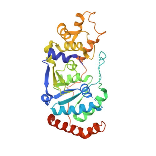
    5FV7

  • PubMed Abstract: 

    The structure-specific nuclease human flap endonuclease-1 (hFEN1) plays a key role in DNA replication and repair and may be of interest as an oncology target. We present the crystal structure of inhibitor-bound hFEN1, which shows a cyclic N-hydroxyurea bound in the active site coordinated to two magnesium ions. Three such compounds had similar IC50 values but differed subtly in mode of action. One had comparable affinity for protein and protein-substrate complex and prevented reaction by binding to active site catalytic metal ions, blocking the necessary unpairing of substrate DNA. Other compounds were more competitive with substrate. Cellular thermal shift data showed that both inhibitor types engaged with hFEN1 in cells, and activation of the DNA damage response was evident upon treatment with inhibitors. However, cellular EC50 values were significantly higher than in vitro inhibition constants, and the implications of this for exploitation of hFEN1 as a drug target are discussed.


  • Organizational Affiliation

    Centre for Chemical Biology, Department of Chemistry, Krebs Institute, University of Sheffield, Sheffield, UK.


Macromolecules
Find similar proteins by:  (by identity cutoff)  |  3D Structure
Entity ID: 1
MoleculeChains Sequence LengthOrganismDetailsImage
FLAP ENDONUCLEASE 1
A, B
353Homo sapiensMutation(s): 0 
EC: 3.1
UniProt & NIH Common Fund Data Resources
Find proteins for P39748 (Homo sapiens)
Explore P39748 
Go to UniProtKB:  P39748
PHAROS:  P39748
GTEx:  ENSG00000168496 
Entity Groups  
Sequence Clusters30% Identity50% Identity70% Identity90% Identity95% Identity100% Identity
UniProt GroupP39748
Sequence Annotations
Expand
  • Reference Sequence
Experimental Data & Validation

Experimental Data

  • Method: X-RAY DIFFRACTION
  • Resolution: 2.84 Å
  • R-Value Free: 0.300 
  • R-Value Work: 0.234 
  • R-Value Observed: 0.237 
  • Space Group: P 1
Unit Cell:
Length ( Å )Angle ( ˚ )
a = 43.339α = 102.06
b = 50.193β = 94.01
c = 66.945γ = 90.74
Software Package:
Software NamePurpose
REFMACrefinement
XDSdata reduction
SCALAdata scaling
PHASERphasing

Structure Validation

View Full Validation Report


Ligand Structure Quality Assessment 


Entry History 

Revision History  (Full details and data files)

  • Version 1.0: 2016-08-17
    Type: Initial release
  • Version 1.1: 2016-08-24
    Changes: Database references
  • Version 1.2: 2016-10-05
    Changes: Database references
  • Version 1.3: 2017-08-23
    Changes: Data collection