5FEB

Crystal structure of the Voltage-gated Sodium Channel Beta 2 subunit extracellular domain


Experimental Data Snapshot

  • Method: X-RAY DIFFRACTION
  • Resolution: 1.35 Å
  • R-Value Free: 0.206 
  • R-Value Work: 0.172 
  • R-Value Observed: 0.174 

wwPDB Validation   3D Report Full Report


This is version 1.3 of the entry. See complete history


Literature

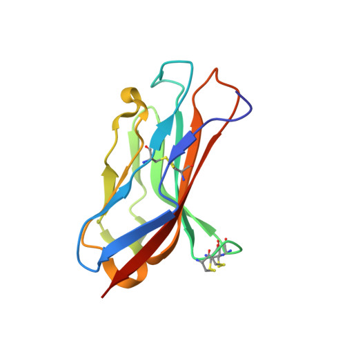
Binary architecture of the Nav1.2-beta 2 signaling complex.

Das, S.Gilchrist, J.Bosmans, F.Van Petegem, F.

(2016) Elife 5

  • DOI: https://doi.org/10.7554/eLife.10960
  • Primary Citation of Related Structures:  
    5FDY, 5FEB

  • PubMed Abstract: 

    To investigate the mechanisms by which β-subunits influence Nav channel function, we solved the crystal structure of the β2 extracellular domain at 1.35Å. We combined these data with known bacterial Nav channel structural insights and novel functional studies to determine the interactions of specific residues in β2 with Nav1.2. We identified a flexible loop formed by (72)Cys and (75)Cys, a unique feature among the four β-subunit isoforms. Moreover, we found that (55)Cys helps to determine the influence of β2 on Nav1.2 toxin susceptibility. Further mutagenesis combined with the use of spider toxins reveals that (55)Cys forms a disulfide bond with (910)Cys in the Nav1.2 domain II pore loop, thereby suggesting a 1:1 stoichiometry. Our results also provide clues as to which disulfide bonds are formed between adjacent Nav1.2 (912/918)Cys residues. The concepts emerging from this work will help to form a model reflecting the β-subunit location in a Nav channel complex.


  • Organizational Affiliation

    Department of Biochemistry and Molecular Biology, Life Sciences Institute, University of British Columbia, Vancouver, Canada.


Macromolecules
Find similar proteins by:  (by identity cutoff)  |  3D Structure
Entity ID: 1
MoleculeChains Sequence LengthOrganismDetailsImage
Sodium channel subunit beta-2125Homo sapiensMutation(s): 1 
Gene Names: SCN2BUNQ326/PRO386
UniProt & NIH Common Fund Data Resources
Find proteins for O60939 (Homo sapiens)
Explore O60939 
Go to UniProtKB:  O60939
PHAROS:  O60939
GTEx:  ENSG00000149575 
Entity Groups  
Sequence Clusters30% Identity50% Identity70% Identity90% Identity95% Identity100% Identity
UniProt GroupO60939
Sequence Annotations
Expand
  • Reference Sequence
Small Molecules
Ligands 1 Unique
IDChains Name / Formula / InChI Key2D Diagram3D Interactions
GOL
Query on GOL

Download Ideal Coordinates CCD File 
B [auth A]GLYCEROL
C3 H8 O3
PEDCQBHIVMGVHV-UHFFFAOYSA-N
Experimental Data & Validation

Experimental Data

  • Method: X-RAY DIFFRACTION
  • Resolution: 1.35 Å
  • R-Value Free: 0.206 
  • R-Value Work: 0.172 
  • R-Value Observed: 0.174 
  • Space Group: P 21 21 21
Unit Cell:
Length ( Å )Angle ( ˚ )
a = 28.7α = 90
b = 59.42β = 90
c = 71.26γ = 90
Software Package:
Software NamePurpose
PHENIXrefinement
HKL-2000data reduction
HKL-2000data scaling
PHASERphasing

Structure Validation

View Full Validation Report


Entry History 

Deposition Data

Revision History  (Full details and data files)

  • Version 1.0: 2016-03-09
    Type: Initial release
  • Version 1.1: 2016-06-22
    Changes: Structure summary
  • Version 1.2: 2017-09-27
    Changes: Author supporting evidence, Derived calculations
  • Version 1.3: 2023-09-27
    Changes: Data collection, Database references, Refinement description