The Human Centriolar Protein CEP135 Contains a Two-Stranded Coiled-Coil Domain Critical for Microtubule Binding.
Kraatz, S., Guichard, P., Obbineni, J.M., Olieric, N., Hatzopoulos, G.N., Hilbert, M., Sen, I., Missimer, J., Gonczy, P., Steinmetz, M.O.(2016) Structure 24: 1358-1371
- PubMed: 27477386
- DOI: https://doi.org/10.1016/j.str.2016.06.011
- Primary Citation of Related Structures:
5FCM, 5FCN - PubMed Abstract:
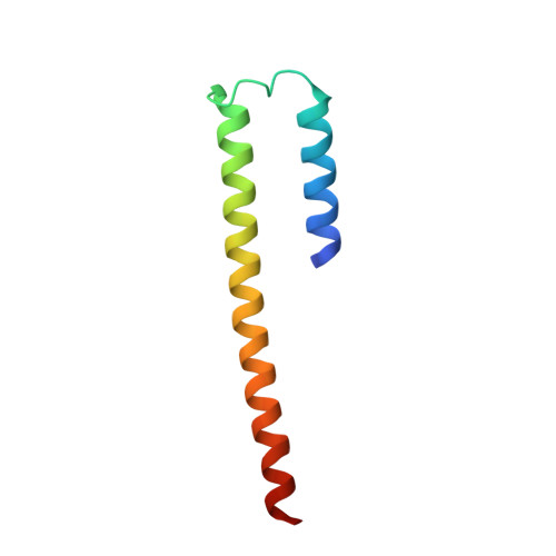
Centrioles are microtubule-based structures that play important roles notably in cell division and cilium biogenesis. CEP135/Bld10p family members are evolutionarily conserved microtubule-binding proteins important for centriole formation. Here, we analyzed in detail the microtubule-binding activity of human CEP135 (HsCEP135). X-ray crystallography and small-angle X-ray scattering in combination with molecular modeling revealed that the 158 N-terminal residues of HsCEP135 (HsCEP135-N) form a parallel two-stranded coiled-coil structure. Biochemical, cryo-electron, and fluorescence microscopy analyses revealed that in vitro HsCEP135-N interacts with tubulin, protofilaments, and microtubules and induces the formation of microtubule bundles. We further identified a 13 amino acid segment spanning residues 96-108, which represents a major microtubule-binding site in HsCEP135-N. Within this segment, we identified a cluster of three lysine residues that contribute to the microtubule bundling activity of HsCEP135-N. Our results provide the first structural information on CEP135/Bld10p proteins and offer insights into their microtubule-binding mechanism.
Organizational Affiliation:
Laboratory of Biomolecular Research, Department of Biology and Chemistry, Paul Scherrer Institut, 5232 Villigen, Switzerland.


