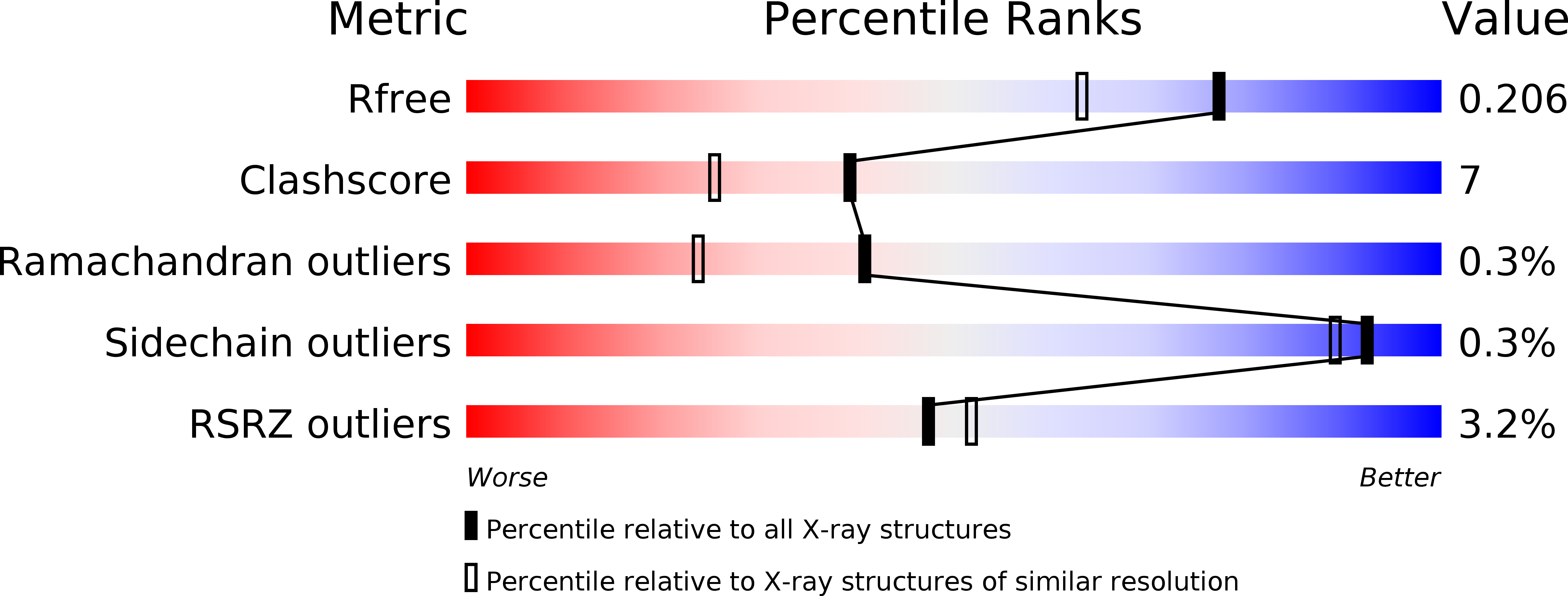
5D3Q

Dynamin 1 GTPase-BSE fusion dimer complexed with GDP


Experimental Data Snapshot

  • Method: X-RAY DIFFRACTION
  • Resolution: 1.70 Å
  • R-Value Free: 0.205 
  • R-Value Work: 0.173 
  • R-Value Observed: 0.174 

wwPDB Validation   3D Report Full Report


Ligand Structure Quality Assessment 


This is version 1.3 of the entry. See complete history


Literature

Crystal structure of the GTPase domain and the bundle signalling element of dynamin in the GDP state.

Anand, R.Eschenburg, S.Reubold, T.F.

(2016) Biochem Biophys Res Commun 469: 76-80

  • DOI: https://doi.org/10.1016/j.bbrc.2015.11.074
  • Primary Citation of Related Structures:  
    5D3Q

  • PubMed Abstract: 

    Dynamin is the prototype of a family of large multi-domain GTPases. The 100 kDa protein is a key player in clathrin-mediated endocytosis, where it cleaves off vesicles from membranes using the energy from GTP hydrolysis. We have solved the high resolution crystal structure of a fusion protein of the GTPase domain and the bundle signalling element (BSE) of dynamin 1 liganded with GDP. The structure provides a hitherto missing snapshot of the GDP state of the hydrolytic cycle of dynamin and reveals how the switch I region moves away from the active site after GTP hydrolysis and release of inorganic phosphate. Comparing our structure of the GDP state with the known structures of the GTP state, the transition state and the nucleotide-free state of dynamin 1 we describe the structural changes through the hydrolytic cycle.


  • Organizational Affiliation

    Hannover Medical School, Institute for Biophysical Chemistry, Carl-Neuberg-Str. 1, 30625 Hannover, Germany.


Macromolecules
Find similar proteins by:  (by identity cutoff)  |  3D Structure
Entity ID: 1
MoleculeChains Sequence LengthOrganismDetailsImage
Dynamin-1,Dynamin-1
A, B
345Homo sapiensMutation(s): 0 
Gene Names: DNM1DNM
EC: 3.6.5.5
UniProt & NIH Common Fund Data Resources
Find proteins for Q05193 (Homo sapiens)
Explore Q05193 
Go to UniProtKB:  Q05193
PHAROS:  Q05193
GTEx:  ENSG00000106976 
Entity Groups  
Sequence Clusters30% Identity50% Identity70% Identity90% Identity95% Identity100% Identity
UniProt GroupQ05193
Sequence Annotations
Expand
  • Reference Sequence
Experimental Data & Validation

Experimental Data

  • Method: X-RAY DIFFRACTION
  • Resolution: 1.70 Å
  • R-Value Free: 0.205 
  • R-Value Work: 0.173 
  • R-Value Observed: 0.174 
  • Space Group: P 1
Unit Cell:
Length ( Å )Angle ( ˚ )
a = 44.22α = 99.4
b = 49.96β = 87.18
c = 82.52γ = 105.18
Software Package:
Software NamePurpose
PHENIXrefinement
XSCALEdata scaling
PHASERphasing
PDB_EXTRACTdata extraction
XDSdata reduction
Cootmodel building

Structure Validation

View Full Validation Report


Ligand Structure Quality Assessment 


Entry History 

Deposition Data

Revision History  (Full details and data files)

  • Version 1.0: 2015-12-02
    Type: Initial release
  • Version 1.1: 2016-09-14
    Changes: Database references
  • Version 1.2: 2017-03-22
    Changes: Data collection
  • Version 1.3: 2024-01-10
    Changes: Data collection, Database references, Refinement description