5CAE

Succinate bound to pig GTP-specific succinyl-CoA synthetase


Experimental Data Snapshot

  • Method: X-RAY DIFFRACTION
  • Resolution: 2.20 Å
  • R-Value Free: 0.214 
  • R-Value Work: 0.165 
  • R-Value Observed: 0.170 

wwPDB Validation   3D Report Full Report


Ligand Structure Quality Assessment 


This is version 1.7 of the entry. See complete history


Literature

Structural basis for the binding of succinate to succinyl-CoA synthetase.

Huang, J.Fraser, M.E.

(2016) Acta Crystallogr D Struct Biol 72: 912-921

  • DOI: https://doi.org/10.1107/S2059798316010044
  • Primary Citation of Related Structures:  
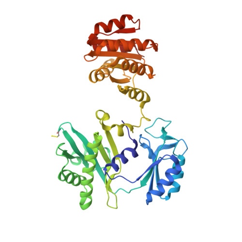
    5CAE

  • PubMed Abstract: 

    Succinyl-CoA synthetase catalyzes the only step in the citric acid cycle that provides substrate-level phosphorylation. Although the binding sites for the substrates CoA, phosphate, and the nucleotides ADP and ATP or GDP and GTP have been identified, the binding site for succinate has not. To determine this binding site, pig GTP-specific succinyl-CoA synthetase was crystallized in the presence of succinate, magnesium ions and CoA, and the structure of the complex was determined by X-ray crystallography to 2.2 Å resolution. Succinate binds in the carboxy-terminal domain of the β-subunit. The succinate-binding site is near both the active-site histidine residue that is phosphorylated in the reaction and the free thiol of CoA. The carboxy-terminal domain rearranges when succinate binds, burying this active site. However, succinate is not in position for transfer of the phosphoryl group from phosphohistidine. Here, it is proposed that when the active-site histidine residue has been phosphorylated by GTP, the phosphohistidine displaces phosphate and triggers the movement of the carboxylate of succinate into position to be phosphorylated. The structure shows why succinyl-CoA synthetase is specific for succinate and does not react appreciably with citrate nor with the other C4-dicarboxylic acids of the citric acid cycle, fumarate and oxaloacetate, but shows some activity with L-malate.


  • Organizational Affiliation

    Department of Biological Sciences, University of Calgary, 2500 University Drive NW, Calgary, Alberta T2N 1N4, Canada.


Macromolecules
Find similar proteins by:  (by identity cutoff)  |  3D Structure
Entity ID: 1
MoleculeChains Sequence LengthOrganismDetailsImage
Succinyl-CoA ligase [ADP/GDP-forming] subunit alpha, mitochondrial313Sus scrofaMutation(s): 0 
Gene Names: SUCLG1
EC: 6.2.1.4 (PDB Primary Data), 6.2.1.5 (PDB Primary Data)
UniProt
Find proteins for O19069 (Sus scrofa)
Explore O19069 
Go to UniProtKB:  O19069
Entity Groups  
Sequence Clusters30% Identity50% Identity70% Identity90% Identity95% Identity100% Identity
UniProt GroupO19069
Sequence Annotations
Expand
  • Reference Sequence
Find similar proteins by:  (by identity cutoff)  |  3D Structure
Entity ID: 2
MoleculeChains Sequence LengthOrganismDetailsImage
Succinyl-CoA ligase [GDP-forming] subunit beta, mitochondrial395Sus scrofaMutation(s): 0 
Gene Names: SUCLG2
EC: 6.2.1.4
UniProt
Find proteins for P53590 (Sus scrofa)
Explore P53590 
Go to UniProtKB:  P53590
Entity Groups  
Sequence Clusters30% Identity50% Identity70% Identity90% Identity95% Identity100% Identity
UniProt GroupP53590
Sequence Annotations
Expand
  • Reference Sequence
Small Molecules
Ligands 6 Unique
IDChains Name / Formula / InChI Key2D Diagram3D Interactions
COA
Query on COA

Download Ideal Coordinates CCD File 
C [auth A]COENZYME A
C21 H36 N7 O16 P3 S
RGJOEKWQDUBAIZ-IBOSZNHHSA-N
SIN
Query on SIN

Download Ideal Coordinates CCD File 
H [auth B]SUCCINIC ACID
C4 H6 O4
KDYFGRWQOYBRFD-UHFFFAOYSA-N
PO4
Query on PO4

Download Ideal Coordinates CCD File 
F [auth A]PHOSPHATE ION
O4 P
NBIIXXVUZAFLBC-UHFFFAOYSA-K
GOL
Query on GOL

Download Ideal Coordinates CCD File 
E [auth A],
I [auth B]
GLYCEROL
C3 H8 O3
PEDCQBHIVMGVHV-UHFFFAOYSA-N
CL
Query on CL

Download Ideal Coordinates CCD File 
D [auth A]CHLORIDE ION
Cl
VEXZGXHMUGYJMC-UHFFFAOYSA-M
MG
Query on MG

Download Ideal Coordinates CCD File 
G [auth B]MAGNESIUM ION
Mg
JLVVSXFLKOJNIY-UHFFFAOYSA-N
Modified Residues  1 Unique
IDChains TypeFormula2D DiagramParent
CSS
Query on CSS
B
L-PEPTIDE LINKINGC3 H7 N O2 S2CYS
Binding Affinity Annotations 
IDSourceBinding Affinity
SIN Binding MOAD:  5CAE Ki: 5.00e+7 (nM) from 1 assay(s)
Experimental Data & Validation

Experimental Data

  • Method: X-RAY DIFFRACTION
  • Resolution: 2.20 Å
  • R-Value Free: 0.214 
  • R-Value Work: 0.165 
  • R-Value Observed: 0.170 
  • Space Group: P 1 21 1
Unit Cell:
Length ( Å )Angle ( ˚ )
a = 86.67α = 90
b = 81.02β = 104.9
c = 50.78γ = 90
Software Package:
Software NamePurpose
PHENIXrefinement
SCALAdata scaling
iMOSFLMdata reduction
Cootmodel building
PHENIXphasing

Structure Validation

View Full Validation Report


Ligand Structure Quality Assessment 


Entry History & Funding Information

Deposition Data


Funding OrganizationLocationGrant Number
Natural Sciences and Engineering Research Council (NSERC, Canada)Canada--

Revision History  (Full details and data files)

  • Version 1.0: 2016-06-29
    Type: Initial release
  • Version 1.1: 2016-08-10
    Changes: Database references
  • Version 1.2: 2016-08-17
    Changes: Database references
  • Version 1.3: 2017-09-06
    Changes: Author supporting evidence, Derived calculations
  • Version 1.4: 2017-11-01
    Changes: Author supporting evidence
  • Version 1.5: 2020-01-08
    Changes: Author supporting evidence
  • Version 1.6: 2023-09-27
    Changes: Data collection, Database references, Derived calculations, Refinement description
  • Version 1.7: 2023-11-15
    Changes: Data collection