4XNU

X-ray structure of Drosophila dopamine transporter in complex with nisoxetine


Experimental Data Snapshot

  • Method: X-RAY DIFFRACTION
  • Resolution: 2.98 Å
  • R-Value Free: 0.251 
  • R-Value Work: 0.226 
  • R-Value Observed: 0.227 

wwPDB Validation   3D Report Full Report


Ligand Structure Quality Assessment 


This is version 1.4 of the entry. See complete history


Literature

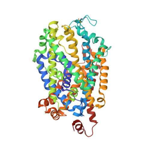
X-ray structures of Drosophila dopamine transporter in complex with nisoxetine and reboxetine.

Penmatsa, A.Wang, K.H.Gouaux, E.

(2015) Nat Struct Mol Biol 22: 506-508

  • DOI: https://doi.org/10.1038/nsmb.3029
  • Primary Citation of Related Structures:  
    4XNU, 4XNX

  • PubMed Abstract: 

    Most antidepressants elicit their therapeutic benefits through selective blockade of Na(+)/Cl(-)-coupled neurotransmitter transporters. Here we report X-ray structures of the Drosophila melanogaster dopamine transporter in complexes with the polycyclic antidepressants nisoxetine or reboxetine. The inhibitors stabilize the transporter in an outward-open conformation by occupying the substrate-binding site. These structures explain how interactions between the binding pocket and substituents on the aromatic rings of antidepressants modulate drug-transporter selectivity.


  • Organizational Affiliation

    Vollum Institute, Oregon Health & Science University, Portland OR.


Macromolecules
Find similar proteins by:  (by identity cutoff)  |  3D Structure
Entity ID: 1
MoleculeChains Sequence LengthOrganismDetailsImage
Integral membrane protein-dopamine transporter532Drosophila melanogasterMutation(s): 0 
Membrane Entity: Yes 
UniProt
Find proteins for Q7K4Y6 (Drosophila melanogaster)
Explore Q7K4Y6 
Go to UniProtKB:  Q7K4Y6
Entity Groups  
Sequence Clusters30% Identity50% Identity70% Identity90% Identity95% Identity100% Identity
UniProt GroupQ7K4Y6
Sequence Annotations
Expand
  • Reference Sequence
Find similar proteins by:  (by identity cutoff)  |  3D Structure
Entity ID: 2
MoleculeChains Sequence LengthOrganismDetailsImage
antibody fragment light chainB [auth L]214Mus musculusMutation(s): 0 
Entity Groups  
Sequence Clusters30% Identity50% Identity70% Identity90% Identity95% Identity100% Identity
Sequence Annotations
Expand
  • Reference Sequence
Find similar proteins by:  (by identity cutoff)  |  3D Structure
Entity ID: 3
MoleculeChains Sequence LengthOrganismDetailsImage
antibody fragment heavy chainC [auth H]240Mus musculusMutation(s): 0 
Entity Groups  
Sequence Clusters30% Identity50% Identity70% Identity90% Identity95% Identity100% Identity
Sequence Annotations
Expand
  • Reference Sequence
Experimental Data & Validation

Experimental Data

  • Method: X-RAY DIFFRACTION
  • Resolution: 2.98 Å
  • R-Value Free: 0.251 
  • R-Value Work: 0.226 
  • R-Value Observed: 0.227 
  • Space Group: P 21 21 21
Unit Cell:
Length ( Å )Angle ( ˚ )
a = 96.901α = 90
b = 133.667β = 90
c = 161.682γ = 90
Software Package:
Software NamePurpose
HKL-2000data reduction
PHENIXrefinement
PDB_EXTRACTdata extraction
SCALEPACKdata scaling
PHASERphasing

Structure Validation

View Full Validation Report


Ligand Structure Quality Assessment 


Entry History 

Deposition Data

Revision History  (Full details and data files)

  • Version 1.0: 2015-05-13
    Type: Initial release
  • Version 1.1: 2015-05-27
    Changes: Database references
  • Version 1.2: 2015-06-10
    Changes: Database references
  • Version 1.3: 2017-05-31
    Changes: Other
  • Version 1.4: 2023-09-27
    Changes: Data collection, Database references, Refinement description