


X-ray crystal structure of thioredoxin from Mycobacterium avium
Lukacs, C.M., Arakaki, T., Don Lorimer, D.D., Edwards, T.E.To be published.
Experimental Data Snapshot
wwPDB Validation 3D Report Full Report

Entity ID: 1 | |||||
|---|---|---|---|---|---|
| Molecule | Chains | Sequence Length | Organism | Details | Image |
| Thioredoxin | 125 | Mycobacterium avium subsp. hominissuis 3388 | Mutation(s): 0 Gene Names: MAV3388_24025 | 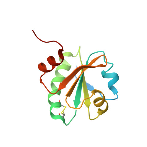 | |
UniProt | |||||
Find proteins for A0A067HWN0 (Mycobacterium avium subsp. hominissuis 3388) Explore A0A067HWN0 Go to UniProtKB: A0A067HWN0 | |||||
Entity Groups | |||||
| Sequence Clusters | 30% Identity50% Identity70% Identity90% Identity95% Identity100% Identity | ||||
| UniProt Group | A0A067HWN0 | ||||
Sequence AnnotationsExpand | |||||
| |||||
| Length ( Å ) | Angle ( ˚ ) |
|---|---|
| a = 26.07 | α = 82.64 |
| b = 29.86 | β = 87.64 |
| c = 31.08 | γ = 66.84 |
| Software Name | Purpose |
|---|---|
| PHENIX | refinement |
| XDS | data reduction |
| XSCALE | data scaling |
RCSB PDB (citation) is hosted by
RCSB PDB is a member of the

