4OVV

Crystal Structure of PI3Kalpha in complex with diC4-PIP2


Experimental Data Snapshot

  • Method: X-RAY DIFFRACTION
  • Resolution: 3.50 Å
  • R-Value Free: 0.339 
  • R-Value Work: 0.237 
  • R-Value Observed: 0.242 

wwPDB Validation   3D Report Full Report


Ligand Structure Quality Assessment 


This is version 1.6 of the entry. See complete history


Literature

Structural basis of nSH2 regulation and lipid binding in PI3K alpha.

Miller, M.S.Schmidt-Kittler, O.Bolduc, D.M.Brower, E.T.Chaves-Moreira, D.Allaire, M.Kinzler, K.W.Jennings, I.G.Thompson, P.E.Cole, P.A.Amzel, L.M.Vogelstein, B.Gabelli, S.B.

(2014) Oncotarget 5: 5198-5208

  • DOI: https://doi.org/10.18632/oncotarget.2263
  • Primary Citation of Related Structures:  
    4OVU, 4OVV

  • PubMed Abstract: 

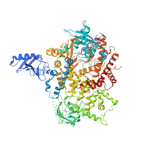
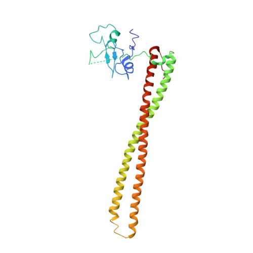
    We report two crystal structures of the wild-type phosphatidylinositol 3-kinase α (PI3Kα) heterodimer refined to 2.9 Å and 3.4 Å resolution: the first as the free enzyme, the second in complex with the lipid substrate, diC4-PIP₂, respectively. The first structure shows key interactions of the N-terminal SH2 domain (nSH2) and iSH2 with the activation loop that suggest a mechanism by which the enzyme is inhibited in its basal state. In the second structure, the lipid substrate binds in a positively charged pocket adjacent to the ATP-binding site, bordered by the P-loop, the activation loop and the iSH2 domain. An additional lipid-binding site was identified at the interface of the ABD, iSH2 and kinase domains. The ability of PI3Kα to bind an additional PIP₂ molecule was confirmed in vitro by fluorescence quenching experiments. The crystal structures reveal key differences in the way the nSH2 domain interacts with wild-type p110α and with the oncogenic mutant p110αH1047R. Increased buried surface area and two unique salt-bridges observed only in the wild-type structure suggest tighter inhibition in the wild-type PI3Kα than in the oncogenic mutant. These differences may be partially responsible for the increased basal lipid kinase activity and increased membrane binding of the oncogenic mutant.


  • Organizational Affiliation

    Medicinal Chemistry, Monash Institute of Pharmaceutical Sciences, Parkville, Victoria, Australia. Present Address: Department of Oncology, Johns Hopkins University School of Medicine, Baltimore Maryland, USA.


Macromolecules
Find similar proteins by:  (by identity cutoff)  |  3D Structure
Entity ID: 1
MoleculeChains Sequence LengthOrganismDetailsImage
Phosphatidylinositol 4,5-bisphosphate 3-kinase catalytic subunit alpha isoform1,096Homo sapiensMutation(s): 0 
Gene Names: PIK3CA
EC: 2.7.1.153
UniProt & NIH Common Fund Data Resources
Find proteins for P42336 (Homo sapiens)
Explore P42336 
Go to UniProtKB:  P42336
PHAROS:  P42336
GTEx:  ENSG00000121879 
Entity Groups  
Sequence Clusters30% Identity50% Identity70% Identity90% Identity95% Identity100% Identity
UniProt GroupP42336
Sequence Annotations
Expand
  • Reference Sequence
Find similar proteins by:  (by identity cutoff)  |  3D Structure
Entity ID: 2
MoleculeChains Sequence LengthOrganismDetailsImage
Phosphatidylinositol 3-kinase regulatory subunit alpha279Homo sapiensMutation(s): 0 
Gene Names: GRB1PI3KR1PIK3R1
UniProt & NIH Common Fund Data Resources
Find proteins for P27986 (Homo sapiens)
Explore P27986 
Go to UniProtKB:  P27986
PHAROS:  P27986
GTEx:  ENSG00000145675 
Entity Groups  
Sequence Clusters30% Identity50% Identity70% Identity90% Identity95% Identity100% Identity
UniProt GroupP27986
Sequence Annotations
Expand
  • Reference Sequence
Small Molecules
Ligands 2 Unique
IDChains Name / Formula / InChI Key2D Diagram3D Interactions
PBU
Query on PBU

Download Ideal Coordinates CCD File 
C [auth A],
E [auth B]
(2R)-3-{[(R)-HYDROXY{[(1R,2R,3S,4R,5R,6S)-2,3,6-TRIHYDROXY-4,5-BIS(PHOSPHONOOXY)CYCLOHEXYL]OXY}PHOSPHORYL]OXY}PROPANE-1 ,2-DIYL DIBUTANOATE
C17 H33 O19 P3
DHAFWWKSHUBGAH-BXJBXBQISA-N
PO4
Query on PO4

Download Ideal Coordinates CCD File 
D [auth A]PHOSPHATE ION
O4 P
NBIIXXVUZAFLBC-UHFFFAOYSA-K
Experimental Data & Validation

Experimental Data

  • Method: X-RAY DIFFRACTION
  • Resolution: 3.50 Å
  • R-Value Free: 0.339 
  • R-Value Work: 0.237 
  • R-Value Observed: 0.242 
  • Space Group: P 21 21 21
Unit Cell:
Length ( Å )Angle ( ˚ )
a = 114.259α = 90
b = 116.083β = 90
c = 148.743γ = 90
Software Package:
Software NamePurpose
REFMACrefinement
PDB_EXTRACTdata extraction
DENZOdata reduction
SCALEPACKdata scaling

Structure Validation

View Full Validation Report


Ligand Structure Quality Assessment 


Entry History & Funding Information

Deposition Data


Funding OrganizationLocationGrant Number
National Institutes of Health/National Cancer Institute (NIH/NCI)United StatesR37 CA043460

Revision History  (Full details and data files)

  • Version 1.0: 2014-09-03
    Type: Initial release
  • Version 1.1: 2014-09-10
    Changes: Structure summary
  • Version 1.2: 2016-08-24
    Changes: Other
  • Version 1.3: 2016-08-31
    Changes: Experimental preparation
  • Version 1.4: 2017-09-27
    Changes: Author supporting evidence, Derived calculations
  • Version 1.5: 2019-12-04
    Changes: Author supporting evidence
  • Version 1.6: 2023-12-27
    Changes: Data collection, Database references, Derived calculations, Structure summary