4LMG

Crystal structure of AFT2 in complex with DNA


Experimental Data Snapshot

  • Method: X-RAY DIFFRACTION
  • Resolution: 2.20 Å
  • R-Value Free: 0.236 
  • R-Value Work: 0.189 
  • R-Value Observed: 0.192 

wwPDB Validation   3D Report Full Report


This is version 1.2 of the entry. See complete history


Literature

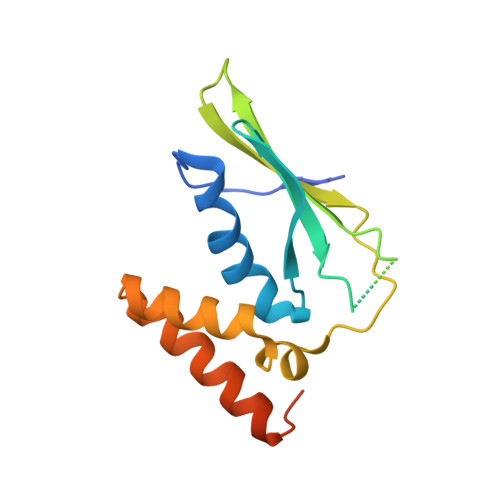
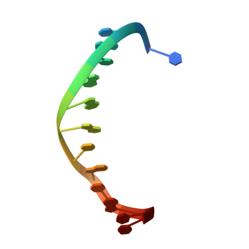
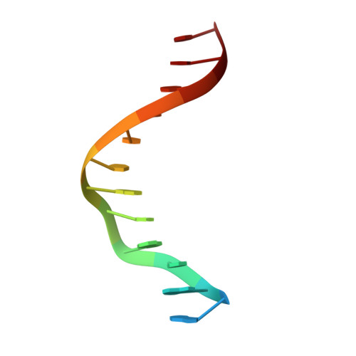
Molecular mechanism and structure of the Saccharomyces cerevisiae iron regulator Aft2.

Poor, C.B.Wegner, S.V.Li, H.Dlouhy, A.C.Schuermann, J.P.Sanishvili, R.Hinshaw, J.R.Riggs-Gelasco, P.J.Outten, C.E.He, C.

(2014) Proc Natl Acad Sci U S A 111: 4043-4048

  • DOI: https://doi.org/10.1073/pnas.1318869111
  • Primary Citation of Related Structures:  
    4LMG

  • PubMed Abstract: 

    The paralogous iron-responsive transcription factors Aft1 and Aft2 (activators of ferrous transport) regulate iron homeostasis in Saccharomyces cerevisiae by activating expression of iron-uptake and -transport genes when intracellular iron is low. We present the previously unidentified crystal structure of Aft2 bound to DNA that reveals the mechanism of DNA recognition via specific interactions of the iron-responsive element with a Zn(2+)-containing WRKY-GCM1 domain in Aft2. We also show that two Aft2 monomers bind a [2Fe-2S] cluster (or Fe(2+)) through a Cys-Asp-Cys motif, leading to dimerization of Aft2 and decreased DNA-binding affinity. Furthermore, we demonstrate that the [2Fe-2S]-bridged heterodimer formed between glutaredoxin-3 and the BolA-like protein Fe repressor of activation-2 transfers a [2Fe-2S] cluster to Aft2 that facilitates Aft2 dimerization. Previous in vivo findings strongly support the [2Fe-2S] cluster-induced dimerization model; however, given the available evidence, Fe(2+)-induced Aft2 dimerization cannot be completely ruled out as an alternative Aft2 inhibition mechanism. Taken together, these data provide insight into the molecular mechanism for iron-dependent transcriptional regulation of Aft2 and highlight the key role of Fe-S clusters as cellular iron signals.


  • Organizational Affiliation

    Department of Chemistry and Institute for Biophysical Dynamics, The University of Chicago, Chicago, IL 60637.


Macromolecules

Find similar proteins by:  (by identity cutoff)  |  3D Structure
Entity ID: 1
MoleculeChains Sequence LengthOrganismDetailsImage
Iron-regulated transcriptional activator AFT2
A, B, C, D
157Saccharomyces cerevisiae S288CMutation(s): 0 
Gene Names: AFT2YPL202C
UniProt
Find proteins for Q08957 (Saccharomyces cerevisiae (strain ATCC 204508 / S288c))
Explore Q08957 
Go to UniProtKB:  Q08957
Entity Groups  
Sequence Clusters30% Identity50% Identity70% Identity90% Identity95% Identity100% Identity
UniProt GroupQ08957
Sequence Annotations
Expand
  • Reference Sequence

Find similar nucleic acids by:  Sequence   |   3D Structure  

Entity ID: 2
MoleculeChains LengthOrganismImage
5'-D(*TP*AP*AP*TP*GP*GP*GP*TP*GP*CP*AP*CP*T)-3'
E, G
13N/A
Sequence Annotations
Expand
  • Reference Sequence

Find similar nucleic acids by:  Sequence   |   3D Structure  

Entity ID: 3
MoleculeChains LengthOrganismImage
5'-D(*AP*AP*GP*TP*GP*CP*AP*CP*CP*CP*AP*TP*T)-3'
F, H
13N/A
Sequence Annotations
Expand
  • Reference Sequence
Experimental Data & Validation

Experimental Data

  • Method: X-RAY DIFFRACTION
  • Resolution: 2.20 Å
  • R-Value Free: 0.236 
  • R-Value Work: 0.189 
  • R-Value Observed: 0.192 
  • Space Group: P 1 21 1
Unit Cell:
Length ( Å )Angle ( ˚ )
a = 41.536α = 90
b = 127.453β = 90.04
c = 70.766γ = 90
Software Package:
Software NamePurpose
XDSdata scaling
PHENIXmodel building
PHENIXrefinement
XDSdata reduction
SCALAdata scaling
PHENIXphasing

Structure Validation

View Full Validation Report


Entry History 

Deposition Data

Revision History  (Full details and data files)

  • Version 1.0: 2014-03-05
    Type: Initial release
  • Version 1.1: 2014-04-23
    Changes: Database references
  • Version 1.2: 2024-02-28
    Changes: Data collection, Database references, Derived calculations