4L0P

Structure of the human EphA3 receptor ligand binding domain complexed with ephrin-A5


Experimental Data Snapshot

  • Method: X-RAY DIFFRACTION
  • Resolution: 2.26 Å
  • R-Value Free: 0.211 
  • R-Value Work: 0.187 
  • R-Value Observed: 0.188 

wwPDB Validation   3D Report Full Report


This is version 2.1 of the entry. See complete history


Literature

Distinctive Structure of the EphA3/Ephrin-A5 Complex Reveals a Dual Mode of Eph Receptor Interaction for Ephrin-A5.

Forse, G.J.Uson, M.L.Nasertorabi, F.Kolatkar, A.Lamberto, I.Pasquale, E.B.Kuhn, P.

(2015) PLoS One 10: e0127081-e0127081

  • DOI: https://doi.org/10.1371/journal.pone.0127081
  • Primary Citation of Related Structures:  
    4L0P

  • PubMed Abstract: 

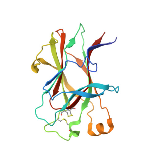
    The Eph receptor tyrosine kinase/ephrin ligand system regulates a wide spectrum of physiological processes, while its dysregulation has been implicated in cancer progression. The human EphA3 receptor is widely upregulated in the tumor microenvironment and is highly expressed in some types of cancer cells. Furthermore, EphA3 is among the most highly mutated genes in lung cancer and it is also frequently mutated in other cancers. We report the structure of the ligand-binding domain of the EphA3 receptor in complex with its preferred ligand, ephrin-A5. The structure of the complex reveals a pronounced tilt of the ephrin-A5 ligand compared to its orientation when bound to the EphA2 and EphB2 receptors and similar to its orientation when bound to EphA4. This tilt brings an additional area of ephrin-A5 into contact with regions of EphA3 outside the ephrin-binding pocket thereby enlarging the size of the interface, which is consistent with the high binding affinity of ephrin-A5 for EphA3. This large variation in the tilt of ephrin-A5 bound to different Eph receptors has not been previously observed for other ephrins.


  • Organizational Affiliation

    Dornsife College of Letters, Arts and Sciences, University of Southern California, 3430 S. Vermont Ave., Suite 105 (110), MC3301, Los Angeles, CA, 90089-3301, United States of America.


Macromolecules
Find similar proteins by:  (by identity cutoff)  |  3D Structure
Entity ID: 1
MoleculeChains Sequence LengthOrganismDetailsImage
Ephrin type-A receptor 3176Homo sapiensMutation(s): 0 
Gene Names: EPHA3EPHA3_HUMANETKETK1HEKTYRO4
UniProt & NIH Common Fund Data Resources
Find proteins for P29320 (Homo sapiens)
Explore P29320 
Go to UniProtKB:  P29320
PHAROS:  P29320
GTEx:  ENSG00000044524 
Entity Groups  
Sequence Clusters30% Identity50% Identity70% Identity90% Identity95% Identity100% Identity
UniProt GroupP29320
Sequence Annotations
Expand
  • Reference Sequence
Find similar proteins by:  (by identity cutoff)  |  3D Structure
Entity ID: 2
MoleculeChains Sequence LengthOrganismDetailsImage
Ephrin-A5143Homo sapiensMutation(s): 0 
Gene Names: EFNA5EFNA5_HUMANEPLG7LERK7
UniProt & NIH Common Fund Data Resources
Find proteins for P52803 (Homo sapiens)
Explore P52803 
Go to UniProtKB:  P52803
PHAROS:  P52803
GTEx:  ENSG00000184349 
Entity Groups  
Sequence Clusters30% Identity50% Identity70% Identity90% Identity95% Identity100% Identity
UniProt GroupP52803
Sequence Annotations
Expand
  • Reference Sequence
Oligosaccharides

Help

Entity ID: 3
MoleculeChains Length2D Diagram Glycosylation3D Interactions
2-acetamido-2-deoxy-beta-D-glucopyranose-(1-4)-2-acetamido-2-deoxy-beta-D-glucopyranose
C
2N-Glycosylation
Glycosylation Resources
GlyTouCan:  G42666HT
GlyCosmos:  G42666HT
GlyGen:  G42666HT
Experimental Data & Validation

Experimental Data

  • Method: X-RAY DIFFRACTION
  • Resolution: 2.26 Å
  • R-Value Free: 0.211 
  • R-Value Work: 0.187 
  • R-Value Observed: 0.188 
  • Space Group: P 43
Unit Cell:
Length ( Å )Angle ( ˚ )
a = 60.043α = 90
b = 60.043β = 90
c = 91.574γ = 90
Software Package:
Software NamePurpose
Blu-Icedata collection
AutoSolphasing
PHENIXrefinement
HKL-2000data reduction
HKL-2000data scaling

Structure Validation

View Full Validation Report


Entry History 

Deposition Data

Revision History  (Full details and data files)

  • Version 1.0: 2014-06-11
    Type: Initial release
  • Version 1.1: 2015-04-01
    Changes: Other
  • Version 1.2: 2015-07-01
    Changes: Database references
  • Version 1.3: 2017-11-15
    Changes: Refinement description
  • Version 2.0: 2020-07-29
    Type: Remediation
    Reason: Carbohydrate remediation
    Changes: Atomic model, Data collection, Database references, Derived calculations, Structure summary
  • Version 2.1: 2023-09-20
    Changes: Data collection, Database references, Refinement description, Structure summary