Structural, kinetic and computational investigation of Vitis vinifera DHDPS reveals new insight into the mechanism of lysine-mediated allosteric inhibition.
Atkinson, S.C., Dogovski, C., Downton, M.T., Czabotar, P.E., Dobson, R.C., Gerrard, J.A., Wagner, J., Perugini, M.A.(2013) Plant Mol Biol 81: 431-446
- PubMed: 23354837
- DOI: https://doi.org/10.1007/s11103-013-0014-7
- Primary Citation of Related Structures:
4HNN - PubMed Abstract:
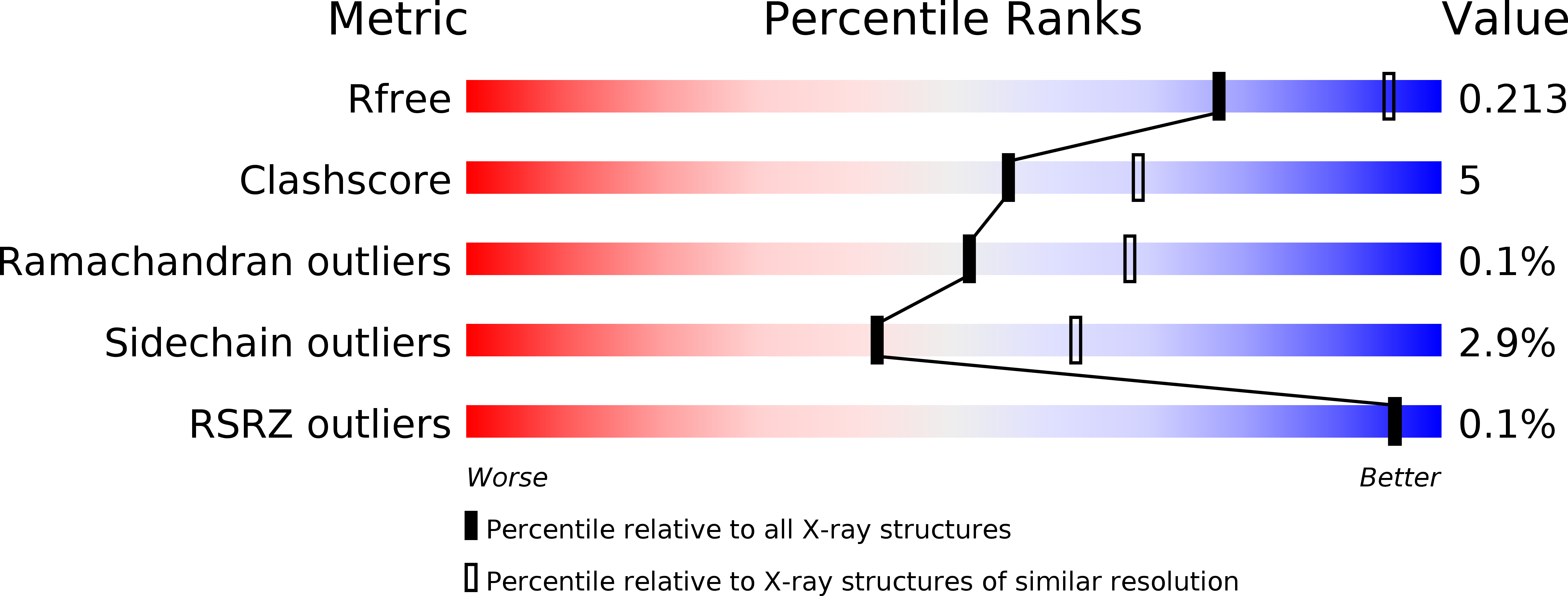
Lysine is one of the most limiting amino acids in plants and its biosynthesis is carefully regulated through inhibition of the first committed step in the pathway catalyzed by dihydrodipicolinate synthase (DHDPS). This is mediated via a feedback mechanism involving the binding of lysine to the allosteric cleft of DHDPS. However, the precise allosteric mechanism is yet to be defined. We present a thorough enzyme kinetic and thermodynamic analysis of lysine inhibition of DHDPS from the common grapevine, Vitis vinifera (Vv). Our studies demonstrate that lysine binding is both tight (relative to bacterial DHDPS orthologs) and cooperative. The crystal structure of the enzyme bound to lysine (2.4 Å) identifies the allosteric binding site and clearly shows a conformational change of several residues within the allosteric and active sites. Molecular dynamics simulations comparing the lysine-bound (PDB ID 4HNN) and lysine free (PDB ID 3TUU) structures show that Tyr132, a key catalytic site residue, undergoes significant rotational motion upon lysine binding. This suggests proton relay through the catalytic triad is attenuated in the presence of lysine. Our study reveals for the first time the structural mechanism for allosteric inhibition of DHDPS from the common grapevine.
Organizational Affiliation:
Department of Biochemistry, La Trobe Institute for Molecular Science, La Trobe University, Melbourne, VIC, 3086, Australia.


