4EB1

Hyperstable in-frame insertion variant of antithrombin


Experimental Data Snapshot

  • Method: X-RAY DIFFRACTION
  • Resolution: 2.80 Å
  • R-Value Free: 0.255 
  • R-Value Work: 0.198 
  • R-Value Observed: 0.201 

wwPDB Validation   3D Report Full Report


Ligand Structure Quality Assessment 


This is version 2.1 of the entry. See complete history


Literature

Type II antithrombin deficiency caused by a large in-frame insertion: structural, functional and pathological relevance.

Martinez-Martinez, I.Johnson, D.J.Yamasaki, M.Navarro-Fernandez, J.Ordonez, A.Vicente, V.Huntington, J.A.Corral, J.

(2012) J Thromb Haemost 10: 1859-1866

  • DOI: https://doi.org/10.1111/j.1538-7836.2012.04839.x
  • Primary Citation of Related Structures:  
    4EB1

  • PubMed Abstract: 

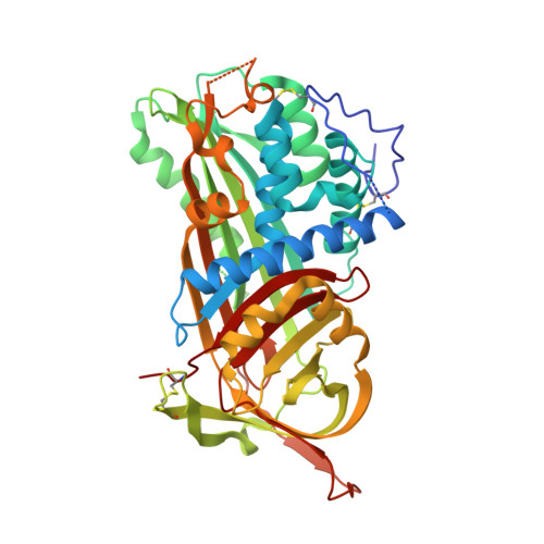
    The metastable native conformation of serpins is required for their protease inhibition mechanism, but also renders them vulnerable to missense mutations that promote protein misfolding with pathological consequences. To characterize the first antithrombin deficiency caused by a large in-frame insertion. Functional, biochemical and molecular analysis of the proband and relatives was performed. Recombinant antithrombin was expressed in HEK-EBNA cells. Plasma and recombinant antithrombins were purified and sequenced by Edman degradation. The stability was evaluated by calorimetry. Reactive centre loop (RCL) exposure was determined by thrombin cleavage. Mutant antithrombin was crystallized as a dimer with latent plasma antithrombin. The patient, with a spontaneous pulmonary embolism, belongs to a family with significant thrombotic history. We identified a complex heterozygous in-frame insertion of 24 bp in SERPINC1, affecting strand 3 of β-sheet A, a region highly conserved in serpins. Surprisingly, the insertion resulted in a type II antithrombin deficiency with heparin binding defect. The mutant antithrombin, with a molecular weight of 59 kDa, had a proteolytic cleavage at W49 but maintained the N-terminal disulphide bonds, and was conformationally sensitive. The variant was non-inhibitory. Analysis of the crystal structure of the hyperstable recombinant protein showed that the inserted sequence annealed into β-sheet A as the fourth strand, and maintained a native RCL. This is the first case of a large in frame-insertion that allows correct folding, glycosylation, and secretion of a serpin, resulting in a conformationally sensitive non-inhibitory variant, which acquires a hyperstable conformation with a native RCL.


  • Organizational Affiliation

    Centro Regional de Hemodonación, University of Murcia, Regional Campus of International Excellence Campus Mare Nostrum, Murcia, Spain.


Macromolecules
Find similar proteins by:  (by identity cutoff)  |  3D Structure
Entity ID: 1
MoleculeChains Sequence LengthOrganismDetailsImage
Antithrombin-IIIA [auth I]440Homo sapiensMutation(s): 1 
Gene Names: SERPINC1AT3PRO0309
UniProt & NIH Common Fund Data Resources
Find proteins for P01008 (Homo sapiens)
Explore P01008 
Go to UniProtKB:  P01008
PHAROS:  P01008
GTEx:  ENSG00000117601 
Entity Groups  
Sequence Clusters30% Identity50% Identity70% Identity90% Identity95% Identity100% Identity
UniProt GroupP01008
Sequence Annotations
Expand
  • Reference Sequence
Find similar proteins by:  (by identity cutoff)  |  3D Structure
Entity ID: 2
MoleculeChains Sequence LengthOrganismDetailsImage
Antithrombin-IIIB [auth L]432Homo sapiensMutation(s): 0 
UniProt & NIH Common Fund Data Resources
Find proteins for P01008 (Homo sapiens)
Explore P01008 
Go to UniProtKB:  P01008
PHAROS:  P01008
GTEx:  ENSG00000117601 
Entity Groups  
Sequence Clusters30% Identity50% Identity70% Identity90% Identity95% Identity100% Identity
UniProt GroupP01008
Sequence Annotations
Expand
  • Reference Sequence
Oligosaccharides

Help

Entity ID: 3
MoleculeChains Length2D Diagram Glycosylation3D Interactions
2-acetamido-2-deoxy-beta-D-glucopyranose-(1-4)-2-acetamido-2-deoxy-beta-D-glucopyranoseC [auth A],
D [auth B]
2N-Glycosylation
Glycosylation Resources
GlyTouCan:  G42666HT
GlyCosmos:  G42666HT
GlyGen:  G42666HT
Small Molecules
Ligands 1 Unique
IDChains Name / Formula / InChI Key2D Diagram3D Interactions
NAG
Query on NAG

Download Ideal Coordinates CCD File 
E [auth I],
F [auth I]
2-acetamido-2-deoxy-beta-D-glucopyranose
C8 H15 N O6
OVRNDRQMDRJTHS-FMDGEEDCSA-N
Experimental Data & Validation

Experimental Data

  • Method: X-RAY DIFFRACTION
  • Resolution: 2.80 Å
  • R-Value Free: 0.255 
  • R-Value Work: 0.198 
  • R-Value Observed: 0.201 
  • Space Group: P 1 21 1
Unit Cell:
Length ( Å )Angle ( ˚ )
a = 69.75α = 90
b = 101.75β = 105.42
c = 88.81γ = 90
Software Package:
Software NamePurpose
HKL-2000data collection
PHASERphasing
REFMACrefinement
MOSFLMdata reduction
SCALAdata scaling

Structure Validation

View Full Validation Report


Ligand Structure Quality Assessment 


Entry History 

Deposition Data

Revision History  (Full details and data files)

  • Version 1.0: 2012-07-18
    Type: Initial release
  • Version 1.1: 2012-09-19
    Changes: Database references
  • Version 1.2: 2017-07-26
    Changes: Refinement description, Source and taxonomy
  • Version 2.0: 2020-07-29
    Type: Remediation
    Reason: Carbohydrate remediation
    Changes: Atomic model, Data collection, Database references, Derived calculations, Structure summary
  • Version 2.1: 2023-09-13
    Changes: Data collection, Database references, Refinement description, Structure summary