4BE4

Closed conformation of O. piceae sterol esterase


Experimental Data Snapshot

  • Method: X-RAY DIFFRACTION
  • Resolution: 2.60 Å
  • R-Value Free: 0.221 
  • R-Value Work: 0.164 
  • R-Value Observed: 0.167 

wwPDB Validation   3D Report Full Report


Ligand Structure Quality Assessment 


This is version 1.2 of the entry. See complete history


Literature

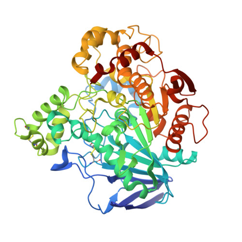
Crystal Structures of Ophiostoma Piceae Sterol Esterase: Structural Insights Into Activation Mechanism and Product Release.

Gutierrez-Fernandez, J.Vaquero, M.E.Prieto, A.Barriuso, J.Martinez, M.J.Hermoso, J.A.

(2014) J Struct Biol 187: 215

  • DOI: https://doi.org/10.1016/j.jsb.2014.07.007
  • Primary Citation of Related Structures:  
    4BE4, 4BE9, 4UPD

  • PubMed Abstract: 

    Sterol esterases are able to efficiently hydrolyze both sterol esters and triglycerides and to carry out synthesis reactions in the presence of organic solvents. Their high versatility makes them excellent candidates for biotechnological purposes. Sterol esterase from fungus Ophiostoma piceae (OPE) belongs to the family abH03.01 of the Candida rugosa lipase-like proteins. Crystal structures of OPE were solved in this study for the closed and open conformations. Enzyme activation involves a large displacement of the conserved lid, structural rearrangements of loop α16-α17, and formation of a dimer with a large opening. Three PEG molecules are placed in the active site, mimicking chains of the triglyceride substrate, demonstrating the position of the oxyanion hole and the three pockets that accommodate the sn-1, sn-2 and sn-3 fatty acids chains. One of them is an internal tunnel, connecting the active center with the outer surface of the enzyme 30 Å far from the catalytic Ser220. Based on our structural and biochemical results we propose a mechanism by which a great variety of different substrates can be hydrolyzed in OPE paving the way for the construction of new variants to improve the catalytic properties of these enzymes and their biotechnological applications.


  • Organizational Affiliation

    Departamento de Cristalografía y Biología Estructural, Instituto de Química-Física "Rocasolano", CSIC, Serrano 119, 28006 Madrid, Spain.


Macromolecules
Find similar proteins by:  (by identity cutoff)  |  3D Structure
Entity ID: 1
MoleculeChains Sequence LengthOrganismDetailsImage
STEROL ESTERASE545Ophiostoma piceaeMutation(s): 0 
EC: 3.1.1.13
UniProt
Find proteins for Q2TFW1 (Ophiostoma piceae)
Explore Q2TFW1 
Go to UniProtKB:  Q2TFW1
Entity Groups  
Sequence Clusters30% Identity50% Identity70% Identity90% Identity95% Identity100% Identity
UniProt GroupQ2TFW1
Sequence Annotations
Expand
  • Reference Sequence
Small Molecules
Ligands 5 Unique
IDChains Name / Formula / InChI Key2D Diagram3D Interactions
NAG
Query on NAG

Download Ideal Coordinates CCD File 
B [auth A],
C [auth A]
2-acetamido-2-deoxy-beta-D-glucopyranose
C8 H15 N O6
OVRNDRQMDRJTHS-FMDGEEDCSA-N
MES
Query on MES

Download Ideal Coordinates CCD File 
J [auth A]2-(N-MORPHOLINO)-ETHANESULFONIC ACID
C6 H13 N O4 S
SXGZJKUKBWWHRA-UHFFFAOYSA-N
SO4
Query on SO4

Download Ideal Coordinates CCD File 
Q [auth A],
R [auth A],
S [auth A]
SULFATE ION
O4 S
QAOWNCQODCNURD-UHFFFAOYSA-L
GOL
Query on GOL

Download Ideal Coordinates CCD File 
K [auth A]
L [auth A]
M [auth A]
N [auth A]
O [auth A]
K [auth A],
L [auth A],
M [auth A],
N [auth A],
O [auth A],
P [auth A]
GLYCEROL
C3 H8 O3
PEDCQBHIVMGVHV-UHFFFAOYSA-N
DIO
Query on DIO

Download Ideal Coordinates CCD File 
D [auth A]
E [auth A]
F [auth A]
G [auth A]
H [auth A]
D [auth A],
E [auth A],
F [auth A],
G [auth A],
H [auth A],
I [auth A]
1,4-DIETHYLENE DIOXIDE
C4 H8 O2
RYHBNJHYFVUHQT-UHFFFAOYSA-N
Experimental Data & Validation

Experimental Data

  • Method: X-RAY DIFFRACTION
  • Resolution: 2.60 Å
  • R-Value Free: 0.221 
  • R-Value Work: 0.164 
  • R-Value Observed: 0.167 
  • Space Group: P 63 2 2
Unit Cell:
Length ( Å )Angle ( ˚ )
a = 119.274α = 90
b = 119.274β = 90
c = 206.774γ = 120
Software Package:
Software NamePurpose
PHENIXrefinement
XDSdata reduction
SCALAdata scaling
PHASERphasing

Structure Validation

View Full Validation Report


Ligand Structure Quality Assessment 


Entry History 

Deposition Data

Revision History  (Full details and data files)

  • Version 1.0: 2014-03-19
    Type: Initial release
  • Version 1.1: 2014-09-24
    Changes: Database references
  • Version 1.2: 2020-07-29
    Type: Remediation
    Reason: Carbohydrate remediation
    Changes: Data collection, Derived calculations, Other, Structure summary