4BDX

The structure of the FnI-EGF tandem domain of coagulation factor XII


Experimental Data Snapshot

  • Method: X-RAY DIFFRACTION
  • Resolution: 1.62 Å
  • R-Value Free: 0.247 
  • R-Value Work: 0.216 
  • R-Value Observed: 0.217 

wwPDB Validation   3D Report Full Report


This is version 1.4 of the entry. See complete history


Literature

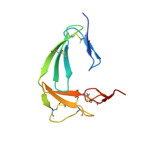
The Structure of the Fni-Egf-Like Tandem Domain of Coagulation Factor Xii Solved Using Siras.

Beringer, D.X.Kroon-Batenburg, L.M.J.

(2013) Acta Crystallogr Sect F Struct Biol Cryst Commun 69: 94

  • DOI: https://doi.org/10.1107/S1744309113000286
  • Primary Citation of Related Structures:  
    4BDW, 4BDX

  • PubMed Abstract: 

    Coagulation factor XII (FXII) is a key protein in the intrinsic coagulation and kallikrein-kinin pathways. It has been found that negative surfaces and amyloids, such as Aβ fibrils, can activate FXII. Additionally, it has been suggested that FXII simulates cells and that it plays an important role in thrombosis. To date, no structural data on FXII have been deposited, which makes it difficult to support any hypothesis on the mechanism of FXII function. The crystal structure of the FnI-EGF-like tandem domain of FXII presented here was solved using experimental phases. To determine the phases, a SIRAS approach was used with a native and a holmium chloride-soaked data set. The holmium cluster was coordinated by the C-terminal tails of two symmetry-related molecules. Another observation was that the FnI domain was much more ordered than the EGF-like domain owing to crystal packing. Furthermore, the structure shows the same domain orientation as the homologous FnI-EGF-like tandem domain of tPA. The plausibility of several proposed interactions of these domains of FXII is discussed. Based on this FXII FnI-EGF-like structure, it could be possible that FXII binding to amyloid and negatively charged surfaces is mediated via this part of FXII.


  • Organizational Affiliation

    Crystal and Structural Chemistry, Bijvoet Center for Biomolecular Research, Department of Chemistry, Utrecht University, Padualaan 8, 3584 CH Utrecht, The Netherlands.


Macromolecules
Find similar proteins by:  (by identity cutoff)  |  3D Structure
Entity ID: 1
MoleculeChains Sequence LengthOrganismDetailsImage
COAGULATION FACTOR XIIA HEAVY CHAIN85Homo sapiensMutation(s): 1 
EC: 3.4.21.38
UniProt & NIH Common Fund Data Resources
Find proteins for P00748 (Homo sapiens)
Explore P00748 
Go to UniProtKB:  P00748
PHAROS:  P00748
GTEx:  ENSG00000131187 
Entity Groups  
Sequence Clusters30% Identity50% Identity70% Identity90% Identity95% Identity100% Identity
UniProt GroupP00748
Sequence Annotations
Expand
  • Reference Sequence
Small Molecules
Ligands 1 Unique
IDChains Name / Formula / InChI Key2D Diagram3D Interactions
ACT
Query on ACT

Download Ideal Coordinates CCD File 
B [auth A]ACETATE ION
C2 H3 O2
QTBSBXVTEAMEQO-UHFFFAOYSA-M
Experimental Data & Validation

Experimental Data

  • Method: X-RAY DIFFRACTION
  • Resolution: 1.62 Å
  • R-Value Free: 0.247 
  • R-Value Work: 0.216 
  • R-Value Observed: 0.217 
  • Space Group: I 41 2 2
Unit Cell:
Length ( Å )Angle ( ˚ )
a = 96.788α = 90
b = 96.788β = 90
c = 47.477γ = 90
Software Package:
Software NamePurpose
PHENIXrefinement
XDSdata reduction
Aimlessdata scaling
PHENIXphasing

Structure Validation

View Full Validation Report


Entry History 

Deposition Data

Revision History  (Full details and data files)

  • Version 1.0: 2013-02-13
    Type: Initial release
  • Version 1.1: 2013-02-20
    Changes: Database references
  • Version 1.2: 2013-04-17
    Changes: Other, Structure summary
  • Version 1.3: 2016-12-28
    Changes: Database references
  • Version 1.4: 2017-07-12
    Changes: Refinement description