4AVV

Structure of CPHPC bound to Serum Amyloid P Component


Experimental Data Snapshot

  • Method: X-RAY DIFFRACTION
  • Resolution: 1.60 Å
  • R-Value Free: 0.175 
  • R-Value Work: 0.150 
  • R-Value Observed: 0.150 

wwPDB Validation   3D Report Full Report


Ligand Structure Quality Assessment 


This is version 2.1 of the entry. See complete history


Literature

Interaction of Serum Amyloid P Component with Hexanoyl Bis(D-Proline) (Cphpc)

Kolstoe, S.E.Jenvey, M.C.Purvis, A.Light, M.E.Thompson, D.Hughes, P.Pepys, M.B.Wood, S.P.

(2014) Acta Crystallogr D Biol Crystallogr 70: 2232

  • DOI: https://doi.org/10.1107/S1399004714013455
  • Primary Citation of Related Structures:  
    4AVS, 4AVT, 4AVV, 4AYU

  • PubMed Abstract: 

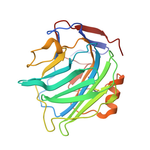
    Under physiological conditions, the pentameric human plasma protein serum amyloid P component (SAP) binds hexanoyl bis(D-proline) (R-1-{6-[R-2-carboxy-pyrrolidin-1-yl]-6-oxo-hexanoyl}pyrrolidine-2-carboxylic acid; CPHPC) through its D-proline head groups in a calcium-dependent interaction. Cooperative effects in binding lead to a substantial enhancement of affinity. Five molecules of the bivalent ligand cross-link and stabilize pairs of SAP molecules, forming a decameric complex that is rapidly cleared from the circulation by the liver. Here, it is reported that X-ray analysis of the SAP complex with CPHPC and cadmium ions provides higher resolution detail of the interaction than is observed with calcium ions. Conformational isomers of CPHPC observed in solution by HPLC and by X-ray analysis are compared with the protein-bound form. These are discussed in relation to the development of CPHPC to provide SAP depletion for the treatment of amyloidosis and other indications.


  • Organizational Affiliation

    Laboratory of Protein Crystallography, Wolfson Drug Discovery Unit, Centre for Amyloidosis and Acute Phase Proteins, Division of Medicine (Royal Free Campus), University College London, Rowland Hill Street, London NW3 2PF, England.


Macromolecules
Find similar proteins by:  (by identity cutoff)  |  3D Structure
Entity ID: 1
MoleculeChains Sequence LengthOrganismDetailsImage
SERUM AMYLOID P-COMPONENT
A, B, C, D, E
204Homo sapiensMutation(s): 0 
UniProt & NIH Common Fund Data Resources
Find proteins for P02743 (Homo sapiens)
Explore P02743 
Go to UniProtKB:  P02743
PHAROS:  P02743
GTEx:  ENSG00000132703 
Entity Groups  
Sequence Clusters30% Identity50% Identity70% Identity90% Identity95% Identity100% Identity
UniProt GroupP02743
Sequence Annotations
Expand
  • Reference Sequence
Oligosaccharides

Help

Entity ID: 2
MoleculeChains Length2D Diagram Glycosylation3D Interactions
2-acetamido-2-deoxy-beta-D-glucopyranose-(1-4)-2-acetamido-2-deoxy-beta-D-glucopyranose
F
2N-Glycosylation
Glycosylation Resources
GlyTouCan:  G42666HT
GlyCosmos:  G42666HT
GlyGen:  G42666HT
Entity ID: 3
MoleculeChains Length2D Diagram Glycosylation3D Interactions
N-acetyl-alpha-neuraminic acid-(2-6)-beta-D-galactopyranose-(1-4)-2-acetamido-2-deoxy-beta-D-glucopyranose-(1-2)-alpha-D-mannopyranose-(1-3)-[N-acetyl-alpha-neuraminic acid-(2-6)-beta-D-galactopyranose-(1-4)-2-acetamido-2-deoxy-beta-D-glucopyranose-(1-2)-alpha-D-mannopyranose-(1-6)]beta-D-mannopyranose-(1-4)-2-acetamido-2-deoxy-beta-D-glucopyranose-(1-4)-2-acetamido-2-deoxy-beta-D-glucopyranose
G
11N-Glycosylation
Glycosylation Resources
GlyTouCan:  G84467IZ
GlyCosmos:  G84467IZ
GlyGen:  G84467IZ
Small Molecules
Ligands 5 Unique
IDChains Name / Formula / InChI Key2D Diagram3D Interactions
GHE
Query on GHE

Download Ideal Coordinates CCD File 
HA [auth C],
J [auth A],
NA [auth D],
UA [auth E],
V [auth B]
(2R)-1-[6-[(2R)-2-carboxypyrrolidin-1-yl]-6-oxidanylidene-hexanoyl]pyrrolidine-2-carboxylic acid
C16 H24 N2 O6
HZLAWYIBLZNRFZ-VXGBXAGGSA-N
SIA
Query on SIA

Download Ideal Coordinates CCD File 
BA [auth B]N-acetyl-alpha-neuraminic acid
C11 H19 N O9
SQVRNKJHWKZAKO-YRMXFSIDSA-N
NAG
Query on NAG

Download Ideal Coordinates CCD File 
AB [auth E],
JA [auth C],
Q [auth A]
2-acetamido-2-deoxy-beta-D-glucopyranose
C8 H15 N O6
OVRNDRQMDRJTHS-FMDGEEDCSA-N
CD
Query on CD

Download Ideal Coordinates CCD File 
AA [auth B]
CA [auth B]
DA [auth B]
DB [auth E]
EA [auth B]
AA [auth B],
CA [auth B],
DA [auth B],
DB [auth E],
EA [auth B],
EB [auth E],
FA [auth C],
FB [auth E],
GA [auth C],
GB [auth E],
IA [auth C],
KA [auth C],
L [auth A],
LA [auth C],
M [auth A],
MA [auth C],
N [auth A],
O [auth A],
OA [auth D],
P [auth A],
PA [auth D],
QA [auth D],
RA [auth D],
S [auth A],
SA [auth D],
T [auth A],
U [auth A],
VA [auth E],
WA [auth E],
X [auth B],
XA [auth E],
Y [auth B],
YA [auth E],
Z [auth B],
ZA [auth E]
CADMIUM ION
Cd
WLZRMCYVCSSEQC-UHFFFAOYSA-N
ACT
Query on ACT

Download Ideal Coordinates CCD File 
BB [auth E]
CB [auth E]
H [auth A]
I [auth A]
K [auth A]
BB [auth E],
CB [auth E],
H [auth A],
I [auth A],
K [auth A],
R [auth A],
TA [auth E],
W [auth B]
ACETATE ION
C2 H3 O2
QTBSBXVTEAMEQO-UHFFFAOYSA-M
Binding Affinity Annotations 
IDSourceBinding Affinity
GHE Binding MOAD:  4AVV Kd: 8.8 (nM) from 1 assay(s)
Experimental Data & Validation

Experimental Data

  • Method: X-RAY DIFFRACTION
  • Resolution: 1.60 Å
  • R-Value Free: 0.175 
  • R-Value Work: 0.150 
  • R-Value Observed: 0.150 
  • Space Group: C 1 2 1
Unit Cell:
Length ( Å )Angle ( ˚ )
a = 154.379α = 90
b = 108.579β = 138.53
c = 120.263γ = 90
Software Package:
Software NamePurpose
PHENIXrefinement
MOLREPphasing

Structure Validation

View Full Validation Report


Ligand Structure Quality Assessment 


Entry History 

Deposition Data

Revision History  (Full details and data files)

  • Version 1.0: 2013-06-19
    Type: Initial release
  • Version 1.1: 2014-08-06
    Changes: Database references
  • Version 1.2: 2014-10-22
    Changes: Database references
  • Version 2.0: 2020-07-29
    Type: Remediation
    Reason: Carbohydrate remediation
    Changes: Atomic model, Data collection, Derived calculations, Other, Structure summary
  • Version 2.1: 2023-12-20
    Changes: Data collection, Database references, Refinement description, Structure summary