4UFM

Mouse Galactocerebrosidase complexed with 1-deoxy-galacto-nojirimycin DGJ


Experimental Data Snapshot

  • Method: X-RAY DIFFRACTION
  • Resolution: 2.40 Å
  • R-Value Free: 0.209 
  • R-Value Work: 0.171 
  • R-Value Observed: 0.173 

wwPDB Validation   3D Report Full Report


Ligand Structure Quality Assessment 


This is version 2.1 of the entry. See complete history


Literature

Azasugar Inhibitors as Pharmacological Chaperones for Krabbe Disease.

Hill, C.H.Viuff, A.H.Spratley, S.J.Salamone, S.Christensen, S.H.Read, R.J.Moriarty, N.W.Jensen, H.H.Deane, J.E.

(2015) Chem Sci 6: 3075

  • DOI: https://doi.org/10.1039/c5sc00754b
  • Primary Citation of Related Structures:  
    4UFH, 4UFI, 4UFJ, 4UFK, 4UFL, 4UFM

  • PubMed Abstract: 

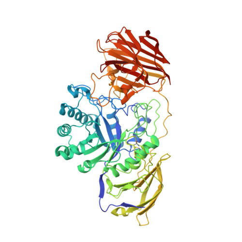
    Krabbe disease is a devastating neurodegenerative disorder characterized by rapid demyelination of nerve fibers. This disease is caused by defects in the lysosomal enzyme β-galactocerebrosidase (GALC), which hydrolyzes the terminal galactose from glycosphingolipids. These lipids are essential components of eukaryotic cell membranes: substrates of GALC include galactocerebroside, the primary lipid component of myelin, and psychosine, a cytotoxic metabolite. Mutations of GALC that cause misfolding of the protein may be responsive to pharmacological chaperone therapy (PCT), whereby small molecules are used to stabilize these mutant proteins, thus correcting trafficking defects and increasing residual catabolic activity in cells. Here we describe a new approach for the synthesis of galacto -configured azasugars and the characterization of their interaction with GALC using biophysical, biochemical and crystallographic methods. We identify that the global stabilization of GALC conferred by azasugar derivatives, measured by fluorescence-based thermal shift assays, is directly related to their binding affinity, measured by enzyme inhibition. X-ray crystal structures of these molecules bound in the GALC active site reveal which residues participate in stabilizing interactions, show how potency is achieved and illustrate the penalties of aza/iminosugar ring distortion. The structure-activity relationships described here identify the key physical properties required of pharmacological chaperones for Krabbe disease and highlight the potential of azasugars as stabilizing agents for future enzyme replacement therapies. This work lays the foundation for new drug-based treatments of Krabbe disease.


  • Organizational Affiliation

    Department of Haematology , Cambridge Institute for Medical Research , University of Cambridge , Cambridge CB2 0XY , UK . Email: jed55@cam.ac.uk.


Macromolecules
Find similar proteins by:  (by identity cutoff)  |  3D Structure
Entity ID: 1
MoleculeChains Sequence LengthOrganismDetailsImage
GALACTOCEREBROSIDASE654Mus musculusMutation(s): 0 
EC: 3.2.1.46
UniProt
Find proteins for P54818 (Mus musculus)
Explore P54818 
Go to UniProtKB:  P54818
Entity Groups  
Sequence Clusters30% Identity50% Identity70% Identity90% Identity95% Identity100% Identity
UniProt GroupP54818
Sequence Annotations
Expand
  • Reference Sequence
Oligosaccharides

Help

Entity ID: 2
MoleculeChains Length2D Diagram Glycosylation3D Interactions
2-acetamido-2-deoxy-beta-D-glucopyranose-(1-4)-2-acetamido-2-deoxy-beta-D-glucopyranose
B, C, D
2N-Glycosylation
Glycosylation Resources
GlyTouCan:  G42666HT
GlyCosmos:  G42666HT
GlyGen:  G42666HT
Small Molecules
Ligands 3 Unique
IDChains Name / Formula / InChI Key2D Diagram3D Interactions
NAG
Query on NAG

Download Ideal Coordinates CCD File 
F [auth A]2-acetamido-2-deoxy-beta-D-glucopyranose
C8 H15 N O6
OVRNDRQMDRJTHS-FMDGEEDCSA-N
DGJ
Query on DGJ

Download Ideal Coordinates CCD File 
E [auth A](2R,3S,4R,5S)-2-(hydroxymethyl)piperidine-3,4,5-triol
C6 H13 N O4
LXBIFEVIBLOUGU-DPYQTVNSSA-N
CA
Query on CA

Download Ideal Coordinates CCD File 
G [auth A]CALCIUM ION
Ca
BHPQYMZQTOCNFJ-UHFFFAOYSA-N
Binding Affinity Annotations 
IDSourceBinding Affinity
DGJ Binding MOAD:  4UFM Ki: 1.90e+5 (nM) from 1 assay(s)
Experimental Data & Validation

Experimental Data

  • Method: X-RAY DIFFRACTION
  • Resolution: 2.40 Å
  • R-Value Free: 0.209 
  • R-Value Work: 0.171 
  • R-Value Observed: 0.173 
  • Space Group: H 3 2
Unit Cell:
Length ( Å )Angle ( ˚ )
a = 248.61α = 90
b = 248.61β = 90
c = 77.54γ = 120
Software Package:
Software NamePurpose
PHENIXrefinement
xia2data reduction
Aimlessdata scaling
PHENIXphasing

Structure Validation

View Full Validation Report


Ligand Structure Quality Assessment 


Entry History 

Deposition Data

Revision History  (Full details and data files)

  • Version 1.0: 2015-04-01
    Type: Initial release
  • Version 1.1: 2015-06-10
    Changes: Database references
  • Version 2.0: 2020-07-29
    Type: Remediation
    Reason: Carbohydrate remediation
    Changes: Atomic model, Data collection, Derived calculations, Other, Structure summary
  • Version 2.1: 2023-12-20
    Changes: Data collection, Database references, Refinement description, Structure summary