4PW5

structure of UHRF2-SRA in complex with a 5hmC-containing DNA, complex I


Experimental Data Snapshot

  • Method: X-RAY DIFFRACTION
  • Resolution: 2.20 Å
  • R-Value Free: 0.206 
  • R-Value Work: 0.161 
  • R-Value Observed: 0.163 

wwPDB Validation   3D Report Full Report


This is version 1.3 of the entry. See complete history


Literature

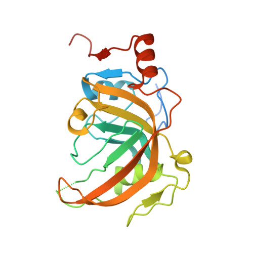
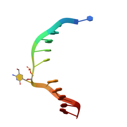
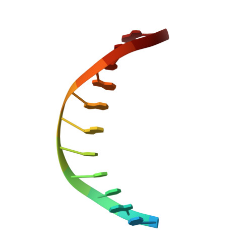
Structural Basis for Hydroxymethylcytosine Recognition by the SRA Domain of UHRF2.

Zhou, T.Xiong, J.Wang, M.Yang, N.Wong, J.Zhu, B.Xu, R.M.

(2014) Mol Cell 54: 879-886

  • DOI: https://doi.org/10.1016/j.molcel.2014.04.003
  • Primary Citation of Related Structures:  
    4PW5, 4PW6, 4PW7

  • PubMed Abstract: 

    Methylated cytosine of CpG dinucleotides in vertebrates may be oxidized by Tet proteins, a process that can lead to DNA demethylation. The predominant oxidation product, 5-hydroxymethylcytosine (5hmC), has been implicated in embryogenesis, cell differentiation, and human diseases. Recently, the SRA domain of UHRF2 (UHRF2-SRA) has been reported to specifically recognize 5hmC, but how UHRF2 recognizes this modification is unclear. Here we report the structure of UHRF2-SRA in complex with a 5hmC-containing DNA. The structure reveals that the conformation of a phenylalanine allows the formation of an optimal 5hmC binding pocket, and a hydrogen bond between the hydroxyl group of 5hmC and UHRF2-SRA is critical for their preferential binding. Further structural and biochemical analyses unveiled the role of SRA domains as a versatile reader of modified DNA, and the knowledge should facilitate further understanding of the biological function of UHRF2 and the comprehension of DNA hydroxymethylation in general.


  • Organizational Affiliation

    National Laboratory of Biomacromolecules, Institute of Biophysics, Chinese Academy of Sciences, Beijing 100101, China; University of Chinese Academy of Sciences, Beijing 100049, China.


Macromolecules

Find similar proteins by:  (by identity cutoff)  |  3D Structure
Entity ID: 1
MoleculeChains Sequence LengthOrganismDetailsImage
E3 ubiquitin-protein ligase UHRF2
A, B, E, F
230Homo sapiensMutation(s): 0 
Gene Names: UHRF2NIRFRNF107
EC: 6.3.2
UniProt & NIH Common Fund Data Resources
Find proteins for Q96PU4 (Homo sapiens)
Explore Q96PU4 
Go to UniProtKB:  Q96PU4
PHAROS:  Q96PU4
GTEx:  ENSG00000147854 
Entity Groups  
Sequence Clusters30% Identity50% Identity70% Identity90% Identity95% Identity100% Identity
UniProt GroupQ96PU4
Sequence Annotations
Expand
  • Reference Sequence

Find similar nucleic acids by:  Sequence   |   3D Structure  

Entity ID: 2
MoleculeChains LengthOrganismImage
5hmC-containing DNA1
C, G
12N/A
Sequence Annotations
Expand
  • Reference Sequence

Find similar nucleic acids by:  Sequence   |   3D Structure  

Entity ID: 3
MoleculeChains LengthOrganismImage
5hmC-containing DNA2
D, H
12N/A
Sequence Annotations
Expand
  • Reference Sequence
Experimental Data & Validation

Experimental Data

  • Method: X-RAY DIFFRACTION
  • Resolution: 2.20 Å
  • R-Value Free: 0.206 
  • R-Value Work: 0.161 
  • R-Value Observed: 0.163 
  • Space Group: P 1
Unit Cell:
Length ( Å )Angle ( ˚ )
a = 42.273α = 90.05
b = 82.807β = 90
c = 82.864γ = 89.98
Software Package:
Software NamePurpose
ADSCdata collection
PHASERphasing
PHENIXrefinement
HKL-2000data reduction
HKL-2000data scaling

Structure Validation

View Full Validation Report


Entry History 

Deposition Data

Revision History  (Full details and data files)

  • Version 1.0: 2014-05-07
    Type: Initial release
  • Version 1.1: 2014-05-21
    Changes: Database references
  • Version 1.2: 2014-06-25
    Changes: Database references
  • Version 1.3: 2023-09-20
    Changes: Data collection, Database references, Derived calculations, Refinement description