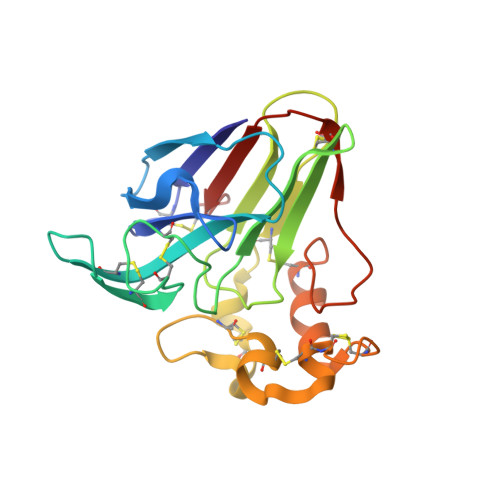
Atomic structure of recombinant thaumatin II reveals flexible conformations in two residues critical for sweetness and three consecutive glycine residues
Masuda, T., Mikami, B., Tani, F.(2014) Biochimie 106: 33-38
- PubMed: 25066915
- DOI: https://doi.org/10.1016/j.biochi.2014.07.016
- Primary Citation of Related Structures:
3WOU - PubMed Abstract:
Thaumatin, an intensely sweet-tasting protein used as a sweetener, elicits a sweet taste at 50 nM. Although two major variants designated thaumatin I and thaumatin II exist in plants, there have been few dedicated thaumatin II structural studies and, to date, data beyond atomic resolution had not been obtained. To identify the detailed structural properties explaining why thaumatin elicits a sweet taste, the structure of recombinant thaumatin II was determined at the resolution of 0.99 Å. Atomic resolution structural analysis with riding hydrogen atoms illustrated the differences in the direction of the side-chains more precisely and the electron density maps of the C-terminal regions were markedly improved. Though it had been suggested that the three consecutive glycine residues (G142-G143-G144) have highly flexible conformations, G143, the central glycine residue was successfully modelled in two conformations for the first time. Furthermore, the side chain r.m.s.d. values for two residues (R67 and R82) critical for sweetness exhibited substantially higher values, suggesting that these residues are highly disordered. These results demonstrated that the flexible conformations in two critical residues favoring their interaction with sweet taste receptors are prominent features of the intensely sweet taste of thaumatin.
Organizational Affiliation:
Division of Food Science and Biotechnology, Graduate School of Agriculture, Kyoto University, Gokasho, Uji, Kyoto 611-0011, Japan. Electronic address: t2masuda@kais.kyoto-u.ac.jp.