3VIV

1510-N membrane-bound stomatin-specific protease K138A mutant in complex with a substrate peptide


Experimental Data Snapshot

  • Method: X-RAY DIFFRACTION
  • Resolution: 2.25 Å
  • R-Value Free: 0.248 
  • R-Value Work: 0.212 
  • R-Value Observed: 0.216 

wwPDB Validation   3D Report Full Report


This is version 1.2 of the entry. See complete history


Literature

Crystal structure of a membrane stomatin-specific protease in complex with a substrate Peptide

Yokoyama, H.Takizawa, N.Kobayashi, D.Matsui, I.Fujii, S.

(2012) Biochemistry 51: 3872-3880

  • DOI: https://doi.org/10.1021/bi300098k
  • Primary Citation of Related Structures:  
    3VIV

  • PubMed Abstract: 

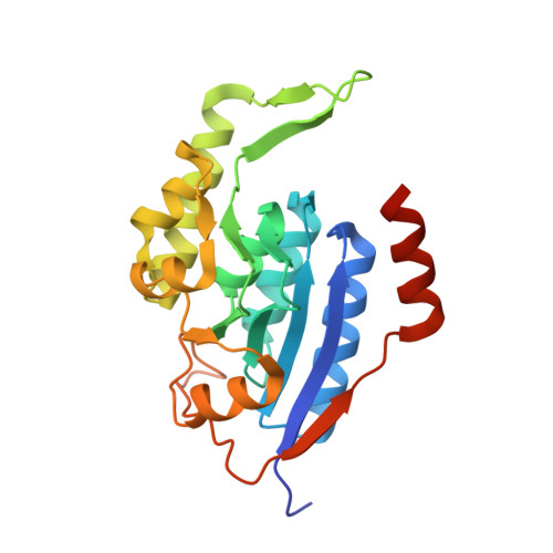
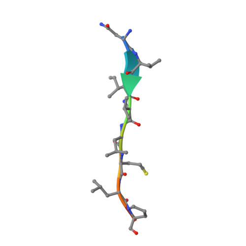
    Membrane-bound proteases are involved in various regulatory functions. A previous report indicated that the N-terminal region of PH1510p (1510-N) from the hyperthermophilic archaeon Pyrococcus horikoshii is a serine protease with a catalytic Ser-Lys dyad (Ser97 and Lys138) and specifically cleaves the C-terminal hydrophobic region of the p-stomatin PH1511p. In humans, an absence of stomatin is associated with a form of hemolytic anemia known as hereditary stomatocytosis. Here, the crystal structure of 1510-N K138A in complex with a peptide substrate was determined at 2.25 Å resolution. In the structure, a 1510-N dimer binds to one peptide. The six central residues (VIVLML) of the peptide are hydrophobic and in a pseudopalindromic structure and therefore favorably fit into the hydrophobic active tunnel of the 1510-N dimer, although 1510-N degrades the substrate at only one point. A comparison with unliganded 1510-N K138A revealed that the binding of the substrate causes a large rotational and translational displacement between protomers and produces a tunnel suitable for binding the peptide. When the peptide binds, the flexible L2 loop of one protomer forms β-strands, whereas that of the other protomer remains in a loop form, indicating that one protomer binds to the peptide more tightly than the other protomer. The Ala138 residues of the two protomers are located very close together (the distance between the two Cβ atoms is 3.6 Å). Thus, in wild-type 1510-N, the close positioning of the catalytic Ser97 and Lys138 residues may be induced by electrostatic repulsion of the two Lys138 side chains of the protomers.


  • Organizational Affiliation

    School of Pharmaceutical Sciences, University of Shizuoka, Shizuoka 422-8526, Japan. h-yokoya@u-shizuoka-ken.ac.jp


Macromolecules
Find similar proteins by:  (by identity cutoff)  |  3D Structure
Entity ID: 1
MoleculeChains Sequence LengthOrganismDetailsImage
441aa long hypothetical nfeD protein
A, B
230Pyrococcus horikoshii OT3Mutation(s): 1 
Gene Names: PH1510
Membrane Entity: Yes 
UniProt
Find proteins for O59179 (Pyrococcus horikoshii (strain ATCC 700860 / DSM 12428 / JCM 9974 / NBRC 100139 / OT-3))
Explore O59179 
Go to UniProtKB:  O59179
Entity Groups  
Sequence Clusters30% Identity50% Identity70% Identity90% Identity95% Identity100% Identity
UniProt GroupO59179
Sequence Annotations
Expand
  • Reference Sequence

Find similar proteins by:  Sequence   |   3D Structure  

Entity ID: 2
MoleculeChains Sequence LengthOrganismDetailsImage
PH STOMATIN PH151110Pyrococcus horikoshii OT3Mutation(s): 0 
Membrane Entity: Yes 
UniProt
Find proteins for O59180 (Pyrococcus horikoshii (strain ATCC 700860 / DSM 12428 / JCM 9974 / NBRC 100139 / OT-3))
Explore O59180 
Go to UniProtKB:  O59180
Entity Groups  
Sequence Clusters30% Identity50% Identity70% Identity90% Identity95% Identity100% Identity
UniProt GroupO59180
Sequence Annotations
Expand
  • Reference Sequence
Experimental Data & Validation

Experimental Data

  • Method: X-RAY DIFFRACTION
  • Resolution: 2.25 Å
  • R-Value Free: 0.248 
  • R-Value Work: 0.212 
  • R-Value Observed: 0.216 
  • Space Group: P 43 21 2
Unit Cell:
Length ( Å )Angle ( ˚ )
a = 111.473α = 90
b = 111.473β = 90
c = 91.768γ = 90
Software Package:
Software NamePurpose
DENZOdata reduction
SCALEPACKdata scaling
MOLREPphasing
REFMACrefinement
PDB_EXTRACTdata extraction
HKL-2000data collection
HKL-2000data reduction
HKL-2000data scaling

Structure Validation

View Full Validation Report


Entry History 

Deposition Data

Revision History  (Full details and data files)

  • Version 1.0: 2012-05-23
    Type: Initial release
  • Version 1.1: 2017-11-22
    Changes: Refinement description
  • Version 1.2: 2023-11-08
    Changes: Data collection, Database references, Derived calculations, Refinement description