3TO9

Crystal structure of yeast Esa1 E338Q HAT domain bound to coenzyme A with active site lysine acetylated


Experimental Data Snapshot

  • Method: X-RAY DIFFRACTION
  • Resolution: 2.00 Å
  • R-Value Free: 0.234 
  • R-Value Work: 0.220 
  • R-Value Observed: 0.220 

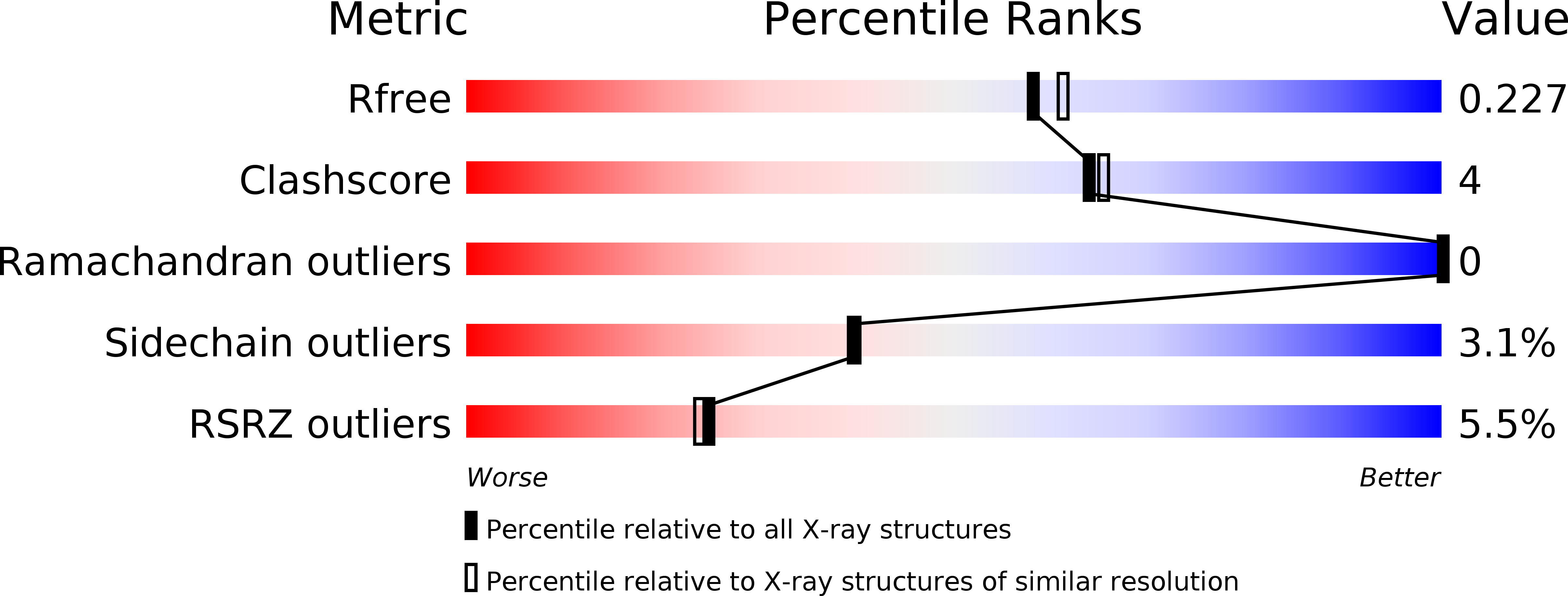
wwPDB Validation   3D Report Full Report


Ligand Structure Quality Assessment 


This is version 1.1 of the entry. See complete history


Literature

MYST protein acetyltransferase activity requires active site lysine autoacetylation.

Yuan, H.Rossetto, D.Mellert, H.Dang, W.Srinivasan, M.Johnson, J.Hodawadekar, S.Ding, E.C.Speicher, K.Abshiru, N.Perry, R.Wu, J.Yang, C.Zheng, Y.G.Speicher, D.W.Thibault, P.Verreault, A.Johnson, F.B.Berger, S.L.Sternglanz, R.McMahon, S.B.Cote, J.Marmorstein, R.

(2011) EMBO J 31: 58-70

  • DOI: https://doi.org/10.1038/emboj.2011.382
  • Primary Citation of Related Structures:  
    3TO6, 3TO7, 3TO9, 3TOA, 3TOB

  • PubMed Abstract: 

    The MYST protein lysine acetyltransferases are evolutionarily conserved throughout eukaryotes and acetylate proteins to regulate diverse biological processes including gene regulation, DNA repair, cell-cycle regulation, stem cell homeostasis and development. Here, we demonstrate that MYST protein acetyltransferase activity requires active site lysine autoacetylation. The X-ray crystal structures of yeast Esa1 (yEsa1/KAT5) bound to a bisubstrate H4K16CoA inhibitor and human MOF (hMOF/KAT8/MYST1) reveal that they are autoacetylated at a strictly conserved lysine residue in MYST proteins (yEsa1-K262 and hMOF-K274) in the enzyme active site. The structure of hMOF also shows partial occupancy of K274 in the unacetylated form, revealing that the side chain reorients to a position that engages the catalytic glutamate residue and would block cognate protein substrate binding. Consistent with the structural findings, we present mass spectrometry data and biochemical experiments to demonstrate that this lysine autoacetylation on yEsa1, hMOF and its yeast orthologue, ySas2 (KAT8) occurs in solution and is required for acetylation and protein substrate binding in vitro. We also show that this autoacetylation occurs in vivo and is required for the cellular functions of these MYST proteins. These findings provide an avenue for the autoposttranslational regulation of MYST proteins that is distinct from other acetyltransferases but draws similarities to the phosphoregulation of protein kinases.


  • Organizational Affiliation

    Gene Expression and Regulation Program, The Wistar Institute, University of Pennsylvania, Philadelphia, PA 19104, USA.


Macromolecules
Find similar proteins by:  (by identity cutoff)  |  3D Structure
Entity ID: 1
MoleculeChains Sequence LengthOrganismDetailsImage
Histone acetyltransferase ESA1276Saccharomyces cerevisiaeMutation(s): 1 
Gene Names: ESA1YOR244WO5257
EC: 2.3.1.48
UniProt
Find proteins for Q08649 (Saccharomyces cerevisiae (strain ATCC 204508 / S288c))
Explore Q08649 
Go to UniProtKB:  Q08649
Entity Groups  
Sequence Clusters30% Identity50% Identity70% Identity90% Identity95% Identity100% Identity
UniProt GroupQ08649
Sequence Annotations
Expand
  • Reference Sequence
Small Molecules
Ligands 4 Unique
IDChains Name / Formula / InChI Key2D Diagram3D Interactions
COA
Query on COA

Download Ideal Coordinates CCD File 
B [auth A]COENZYME A
C21 H36 N7 O16 P3 S
RGJOEKWQDUBAIZ-IBOSZNHHSA-N
CAD
Query on CAD

Download Ideal Coordinates CCD File 
H [auth A]CACODYLIC ACID
C2 H7 As O2
OGGXGZAMXPVRFZ-UHFFFAOYSA-N
SO4
Query on SO4

Download Ideal Coordinates CCD File 
E [auth A],
F [auth A]
SULFATE ION
O4 S
QAOWNCQODCNURD-UHFFFAOYSA-L
EDO
Query on EDO

Download Ideal Coordinates CCD File 
C [auth A],
D [auth A],
G [auth A]
1,2-ETHANEDIOL
C2 H6 O2
LYCAIKOWRPUZTN-UHFFFAOYSA-N
Modified Residues  1 Unique
IDChains TypeFormula2D DiagramParent
ALY
Query on ALY
A
L-PEPTIDE LINKINGC8 H16 N2 O3LYS
Experimental Data & Validation

Experimental Data

  • Method: X-RAY DIFFRACTION
  • Resolution: 2.00 Å
  • R-Value Free: 0.234 
  • R-Value Work: 0.220 
  • R-Value Observed: 0.220 
  • Space Group: I 41 3 2
Unit Cell:
Length ( Å )Angle ( ˚ )
a = 182.47α = 90
b = 182.47β = 90
c = 182.47γ = 90
Software Package:
Software NamePurpose
HKL-2000data collection
PHASERphasing
PHENIXrefinement
HKL-2000data reduction
HKL-2000data scaling

Structure Validation

View Full Validation Report


Ligand Structure Quality Assessment 


Entry History 

Deposition Data

Revision History  (Full details and data files)

  • Version 1.0: 2011-11-09
    Type: Initial release
  • Version 1.1: 2012-01-18
    Changes: Database references