3SSO

MycE Methyltransferase from the Mycinamycin Biosynthetic Pathway in Complex with Mg and SAH, Crystal form 2


Experimental Data Snapshot

  • Method: X-RAY DIFFRACTION
  • Resolution: 1.90 Å
  • R-Value Free: 0.176 
  • R-Value Work: 0.144 
  • R-Value Observed: 0.145 

wwPDB Validation   3D Report Full Report


Ligand Structure Quality Assessment 


This is version 1.4 of the entry. See complete history


Literature

A new structural form in the SAM/metal-dependent o‑methyltransferase family: MycE from the mycinamicin biosynthetic pathway.

Akey, D.L.Li, S.Konwerski, J.R.Confer, L.A.Bernard, S.M.Anzai, Y.Kato, F.Sherman, D.H.Smith, J.L.

(2011) J Mol Biol 413: 438-450

  • DOI: https://doi.org/10.1016/j.jmb.2011.08.040
  • Primary Citation of Related Structures:  
    3SSM, 3SSN, 3SSO

  • PubMed Abstract: 

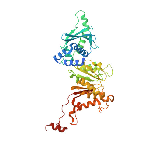
    O-linked methylation of sugar substituents is a common modification in the biosynthesis of many natural products and is catalyzed by multiple families of S-adenosyl-L-methionine (SAM or AdoMet)-dependent methyltransferases (MTs). Mycinamicins, potent antibiotics from Micromonospora griseorubida, can be methylated at two positions on a 6-deoxyallose substituent. The first methylation is catalyzed by MycE, a SAM- and metal-dependent MT. Crystal structures were determined for MycE bound to the product S-adenosyl-L-homocysteine (AdoHcy) and magnesium, both with and without the natural substrate mycinamicin VI. This represents the first structure of a natural product sugar MT in complex with its natural substrate. MycE is a tetramer of a two-domain polypeptide, comprising a C-terminal catalytic MT domain and an N-terminal auxiliary domain, which is important for quaternary assembly and for substrate binding. The symmetric MycE tetramer has a novel MT organization in which each of the four active sites is formed at the junction of three monomers within the tetramer. The active-site structure supports a mechanism in which a conserved histidine acts as a general base, and the metal ion helps to position the methyl acceptor and to stabilize a hydroxylate intermediate. A conserved tyrosine is suggested to support activity through interactions with the transferred methyl group from the SAM methyl donor. The structure of the free enzyme reveals a dramatic order-disorder transition in the active site relative to the S-adenosyl-L-homocysteine complexes, suggesting a mechanism for product/substrate exchange through concerted movement of five loops and the polypeptide C-terminus.


  • Organizational Affiliation

    Life Sciences Institute, University of Michigan, Ann Arbor, MI 48109, USA.


Macromolecules
Find similar proteins by:  (by identity cutoff)  |  3D Structure
Entity ID: 1
MoleculeChains Sequence LengthOrganismDetailsImage
Methyltransferase
A, B, C, D, E
A, B, C, D, E, F
419Micromonospora griseorubidaMutation(s): 0 
Gene Names: mycE
EC: 2.1.1
UniProt
Find proteins for Q83WF2 (Micromonospora griseorubida)
Explore Q83WF2 
Go to UniProtKB:  Q83WF2
Entity Groups  
Sequence Clusters30% Identity50% Identity70% Identity90% Identity95% Identity100% Identity
UniProt GroupQ83WF2
Sequence Annotations
Expand
  • Reference Sequence
Small Molecules
Ligands 2 Unique
IDChains Name / Formula / InChI Key2D Diagram3D Interactions
SAH
Query on SAH

Download Ideal Coordinates CCD File 
H [auth A]
J [auth B]
L [auth C]
N [auth D]
P [auth E]
H [auth A],
J [auth B],
L [auth C],
N [auth D],
P [auth E],
R [auth F]
S-ADENOSYL-L-HOMOCYSTEINE
C14 H20 N6 O5 S
ZJUKTBDSGOFHSH-WFMPWKQPSA-N
MG
Query on MG

Download Ideal Coordinates CCD File 
G [auth A]
I [auth B]
K [auth C]
M [auth D]
O [auth E]
G [auth A],
I [auth B],
K [auth C],
M [auth D],
O [auth E],
Q [auth F]
MAGNESIUM ION
Mg
JLVVSXFLKOJNIY-UHFFFAOYSA-N
Experimental Data & Validation

Experimental Data

  • Method: X-RAY DIFFRACTION
  • Resolution: 1.90 Å
  • R-Value Free: 0.176 
  • R-Value Work: 0.144 
  • R-Value Observed: 0.145 
  • Space Group: C 2 2 21
Unit Cell:
Length ( Å )Angle ( ˚ )
a = 144.22α = 90
b = 250.846β = 90
c = 158.315γ = 90
Software Package:
Software NamePurpose
SCALEPACKdata scaling
PHASERphasing
REFMACrefinement
PDB_EXTRACTdata extraction
HKL-2000data collection
DENZOdata reduction
HKL-2000data scaling

Structure Validation

View Full Validation Report


Ligand Structure Quality Assessment 


Entry History 

Deposition Data

Revision History  (Full details and data files)

  • Version 1.0: 2011-08-24
    Type: Initial release
  • Version 1.1: 2011-12-28
    Changes: Database references
  • Version 1.2: 2014-11-12
    Changes: Structure summary
  • Version 1.3: 2017-11-08
    Changes: Refinement description
  • Version 1.4: 2024-02-28
    Changes: Data collection, Database references, Derived calculations