3QWR

Crystal structure of IL-23 in complex with an adnectin


Experimental Data Snapshot

  • Method: X-RAY DIFFRACTION
  • Resolution: 3.25 Å
  • R-Value Free: 0.264 
  • R-Value Work: 0.234 
  • R-Value Observed: 0.235 

wwPDB Validation   3D Report Full Report


This is version 2.1 of the entry. See complete history


Literature

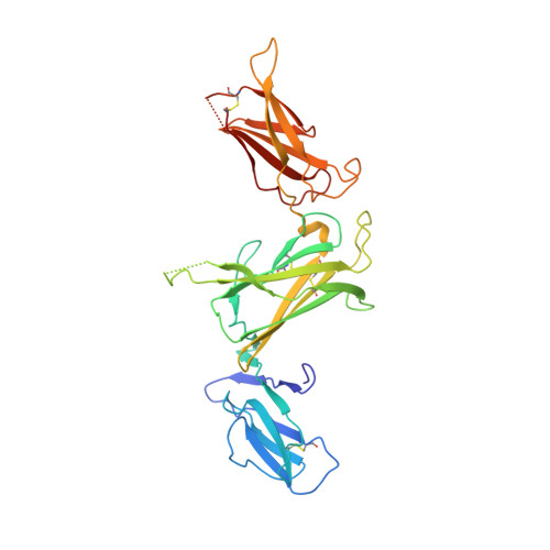
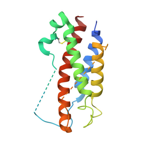
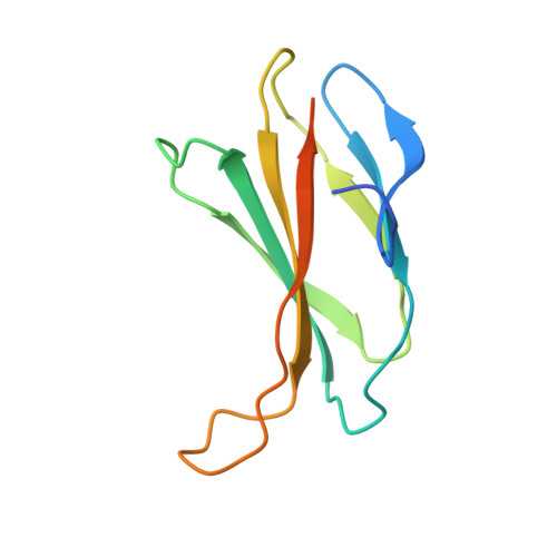
Structures of adnectin/protein complexes reveal an expanded binding footprint.

Ramamurthy, V.Krystek, S.R.Bush, A.Wei, A.Emanuel, S.L.Das Gupta, R.Janjua, A.Cheng, L.Murdock, M.Abramczyk, B.Cohen, D.Lin, Z.Morin, P.Davis, J.H.Dabritz, M.McLaughlin, D.C.Russo, K.A.Chao, G.Wright, M.C.Jenny, V.A.Engle, L.J.Furfine, E.Sheriff, S.

(2012) Structure 20: 259-269

  • DOI: https://doi.org/10.1016/j.str.2011.11.016
  • Primary Citation of Related Structures:  
    3QWQ, 3QWR

  • PubMed Abstract: 

    Adnectins are targeted biologics derived from the tenth type III domain of human fibronectin (¹⁰Fn3), a member of the immunoglobulin superfamily. Target-specific binders are selected from libraries generated by diversifying the three ¹⁰Fn3 loops that are analogous to the complementarity determining regions of antibodies. The crystal structures of two Adnectins were determined, each in complex with its therapeutic target, EGFR or IL-23. Both Adnectins bind different epitopes than those bound by known monoclonal antibodies. Molecular modeling suggests that some of these epitopes might not be accessible to antibodies because of the size and concave shape of the antibody combining site. In addition to interactions from the Adnectin diversified loops, residues from the N terminus and/or the β strands interact with the target proteins in both complexes. Alanine-scanning mutagenesis confirmed the calculated binding energies of these β strand interactions, indicating that these nonloop residues can expand the available binding footprint.


  • Organizational Affiliation

    Bristol-Myers Squibb Research & Development, Princeton, NJ 08543-4000, USA.


Macromolecules
Find similar proteins by:  (by identity cutoff)  |  3D Structure
Entity ID: 1
MoleculeChains Sequence LengthOrganismDetailsImage
Interleukin-12 subunit beta306Homo sapiensMutation(s): 0 
Gene Names: IL12BNKSF2
UniProt & NIH Common Fund Data Resources
Find proteins for P29460 (Homo sapiens)
Explore P29460 
Go to UniProtKB:  P29460
PHAROS:  P29460
GTEx:  ENSG00000113302 
Entity Groups  
Sequence Clusters30% Identity50% Identity70% Identity90% Identity95% Identity100% Identity
UniProt GroupP29460
Sequence Annotations
Expand
  • Reference Sequence
Find similar proteins by:  (by identity cutoff)  |  3D Structure
Entity ID: 2
MoleculeChains Sequence LengthOrganismDetailsImage
Interleukin-23 subunit alpha170Homo sapiensMutation(s): 0 
Gene Names: IL23ASGRFUNQ2498/PRO5798
UniProt & NIH Common Fund Data Resources
Find proteins for Q9NPF7 (Homo sapiens)
Explore Q9NPF7 
Go to UniProtKB:  Q9NPF7
PHAROS:  Q9NPF7
GTEx:  ENSG00000110944 
Entity Groups  
Sequence Clusters30% Identity50% Identity70% Identity90% Identity95% Identity100% Identity
UniProt GroupQ9NPF7
Sequence Annotations
Expand
  • Reference Sequence
Find similar proteins by:  (by identity cutoff)  |  3D Structure
Entity ID: 3
MoleculeChains Sequence LengthOrganismDetailsImage
ADNECTINC [auth D]109Homo sapiensMutation(s): 0 
Entity Groups  
Sequence Clusters30% Identity50% Identity70% Identity90% Identity95% Identity100% Identity
Sequence Annotations
Expand
  • Reference Sequence
Oligosaccharides

Help

Entity ID: 4
MoleculeChains Length2D Diagram Glycosylation3D Interactions
alpha-D-mannopyranose-(1-3)-beta-D-mannopyranose-(1-4)-2-acetamido-2-deoxy-beta-D-glucopyranose-(1-4)-2-acetamido-2-deoxy-beta-D-glucopyranoseD [auth C]4N-Glycosylation
Glycosylation Resources
GlyTouCan:  G81315DD
GlyCosmos:  G81315DD
GlyGen:  G81315DD
Experimental Data & Validation

Experimental Data

  • Method: X-RAY DIFFRACTION
  • Resolution: 3.25 Å
  • R-Value Free: 0.264 
  • R-Value Work: 0.234 
  • R-Value Observed: 0.235 
  • Space Group: I 21 21 21
Unit Cell:
Length ( Å )Angle ( ˚ )
a = 77.7α = 90
b = 91.7β = 90
c = 225.8γ = 90
Software Package:
Software NamePurpose
BUSTER-TNTrefinement
PDB_EXTRACTdata extraction
d*TREKdata reduction
d*TREKdata scaling
PHASERphasing
AMoREphasing
BUSTERrefinement

Structure Validation

View Full Validation Report


Entry History 

Deposition Data

Revision History  (Full details and data files)

  • Version 1.0: 2012-02-01
    Type: Initial release
  • Version 1.1: 2012-02-29
    Changes: Database references
  • Version 1.2: 2017-11-08
    Changes: Refinement description
  • Version 2.0: 2020-07-29
    Type: Remediation
    Reason: Carbohydrate remediation
    Changes: Atomic model, Data collection, Derived calculations, Structure summary
  • Version 2.1: 2023-09-13
    Changes: Data collection, Database references, Refinement description, Structure summary