3MTU

Structure of the Tropomyosin Overlap Complex from Chicken Smooth Muscle


Experimental Data Snapshot

  • Method: X-RAY DIFFRACTION
  • Resolution: 2.10 Å
  • R-Value Free: 0.241 
  • R-Value Work: 0.201 
  • R-Value Observed: 0.203 

wwPDB Validation   3D Report Full Report


This is version 1.3 of the entry. See complete history


Literature

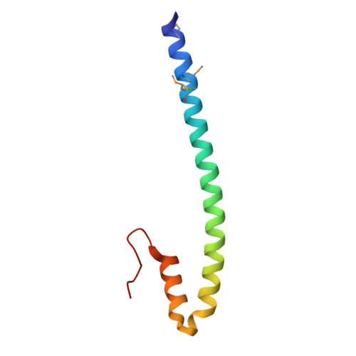
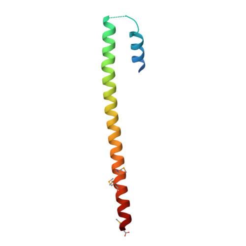
Structure of the tropomyosin overlap complex from chicken smooth muscle: insight into the diversity of N-terminal recognition .

Frye, J.Klenchin, V.A.Rayment, I.

(2010) Biochemistry 49: 4908-4920

  • DOI: https://doi.org/10.1021/bi100349a
  • Primary Citation of Related Structures:  
    3MTU, 3MUD

  • PubMed Abstract: 

    Tropomyosin is a stereotypical alpha-helical coiled coil that polymerizes to form a filamentous macromolecular assembly that lies on the surface of F-actin. The interaction between the C-terminal and N-terminal segments on adjacent molecules is known as the overlap region. We report here two X-ray structures of the chicken smooth muscle tropomyosin overlap complex. A novel approach was used to stabilize the C-terminal and N-terminal fragments. Globular domains from both the human DNA ligase binding protein XRCC4 and bacteriophage varphi29 scaffolding protein Gp7 were fused to 37 and 28 C-terminal amino acid residues of tropomyosin, respectively, whereas the 29 N-terminal amino acids of tropomyosin were fused to the C-terminal helix bundle of microtubule binding protein EB1. The structures of both the XRCC4 and Gp7 fusion proteins complexed with the N-terminal EB1 fusion contain a very similar helix bundle in the overlap region that encompasses approximately 15 residues. The C-terminal coiled coil opens to allow formation of the helix bundle, which is stabilized by hydrophobic interactions. These structures are similar to that observed in the NMR structure of the rat skeletal overlap complex [Greenfield, N. J., et al. (2006) J. Mol. Biol. 364, 80-96]. The interactions between the N- and C-terminal coiled coils of smooth muscle tropomyosin show significant curvature, which differs somewhat between the two structures and implies flexibility in the overlap complex, at least in solution. This is likely an important attribute that allows tropomyosin to assemble around the actin filaments. These structures provide a molecular explanation for the role of N-acetylation in the assembly of native tropomyosin.


  • Organizational Affiliation

    Department of Biochemistry, University of Wisconsin, Madison, Wisconsin 53706, USA.


Macromolecules
Find similar proteins by:  (by identity cutoff)  |  3D Structure
Entity ID: 1
MoleculeChains Sequence LengthOrganismDetailsImage
Tropomyosin alpha-1 chain,Microtubule-associated protein RP/EB family member 1
A, B, C, D
75Gallus gallusHomo sapiens
This entity is chimeric
Mutation(s): 0 
Gene Names: TPM1MAPRE1
UniProt & NIH Common Fund Data Resources
Find proteins for P04268 (Gallus gallus)
Explore P04268 
Go to UniProtKB:  P04268
Find proteins for Q15691 (Homo sapiens)
Explore Q15691 
Go to UniProtKB:  Q15691
PHAROS:  Q15691
GTEx:  ENSG00000101367 
Entity Groups  
Sequence Clusters30% Identity50% Identity70% Identity90% Identity95% Identity100% Identity
UniProt GroupsP04268Q15691
Sequence Annotations
Expand
  • Reference Sequence
Find similar proteins by:  (by identity cutoff)  |  3D Structure
Entity ID: 2
MoleculeChains Sequence LengthOrganismDetailsImage
Capsid assembly scaffolding protein,Tropomyosin alpha-1 chain
E, F
77Salasvirus phi29Gallus gallus
This entity is chimeric
Mutation(s): 0 
UniProt
Find proteins for P04268 (Gallus gallus)
Explore P04268 
Go to UniProtKB:  P04268
Find proteins for P13848 (Bacillus phage phi29)
Explore P13848 
Go to UniProtKB:  P13848
Entity Groups  
Sequence Clusters30% Identity50% Identity70% Identity90% Identity95% Identity100% Identity
UniProt GroupsP04268P13848
Sequence Annotations
Expand
  • Reference Sequence
Small Molecules
Ligands 3 Unique
IDChains Name / Formula / InChI Key2D Diagram3D Interactions
EDO
Query on EDO

Download Ideal Coordinates CCD File 
H [auth A]
I [auth A]
J [auth A]
K [auth A]
P [auth B]
H [auth A],
I [auth A],
J [auth A],
K [auth A],
P [auth B],
S [auth C],
U [auth D],
V [auth D],
X [auth F]
1,2-ETHANEDIOL
C2 H6 O2
LYCAIKOWRPUZTN-UHFFFAOYSA-N
EOH
Query on EOH

Download Ideal Coordinates CCD File 
L [auth A]
M [auth A]
N [auth A]
Q [auth B]
R [auth B]
L [auth A],
M [auth A],
N [auth A],
Q [auth B],
R [auth B],
T [auth C],
W [auth D]
ETHANOL
C2 H6 O
LFQSCWFLJHTTHZ-UHFFFAOYSA-N
CL
Query on CL

Download Ideal Coordinates CCD File 
G [auth A],
O [auth B]
CHLORIDE ION
Cl
VEXZGXHMUGYJMC-UHFFFAOYSA-M
Modified Residues  1 Unique
IDChains TypeFormula2D DiagramParent
MSE
Query on MSE
A, B, C, D
L-PEPTIDE LINKINGC5 H11 N O2 SeMET
Experimental Data & Validation

Experimental Data

  • Method: X-RAY DIFFRACTION
  • Resolution: 2.10 Å
  • R-Value Free: 0.241 
  • R-Value Work: 0.201 
  • R-Value Observed: 0.203 
  • Space Group: C 2 2 2
Unit Cell:
Length ( Å )Angle ( ˚ )
a = 89.131α = 90
b = 285.328β = 90
c = 43.457γ = 90
Software Package:
Software NamePurpose
ADSCdata collection
MLPHAREphasing
REFMACrefinement
HKL-3000data reduction
HKL-2000data scaling

Structure Validation

View Full Validation Report


Entry History 

Deposition Data

Revision History  (Full details and data files)

  • Version 1.0: 2010-06-23
    Type: Initial release
  • Version 1.1: 2011-07-13
    Changes: Version format compliance
  • Version 1.2: 2017-05-31
    Changes: Structure summary
  • Version 1.3: 2018-01-24
    Changes: Structure summary