3MBI

Crystal structure of the phosphoribosylpyrophosphate (PRPP) synthetase from Thermoplasma volcanium in complex with ADP-Mg2+ and ribose 5-phosphate


Experimental Data Snapshot

  • Method: X-RAY DIFFRACTION
  • Resolution: 1.80 Å
  • R-Value Free: 0.219 
  • R-Value Work: 0.176 
  • R-Value Observed: 0.178 

wwPDB Validation   3D Report Full Report


Ligand Structure Quality Assessment 


This is version 2.1 of the entry. See complete history


Literature

The structures of Thermoplasma volcanium phosphoribosyl pyrophosphate synthetase bound to ribose-5-phosphate and ATP analogs.

Cherney, M.M.Cherney, L.T.Garen, C.R.James, M.N.

(2011) J Mol Biol 413: 844-856

  • DOI: https://doi.org/10.1016/j.jmb.2011.09.007
  • Primary Citation of Related Structures:  
    3LPN, 3LRT, 3MBI, 3NAG

  • PubMed Abstract: 

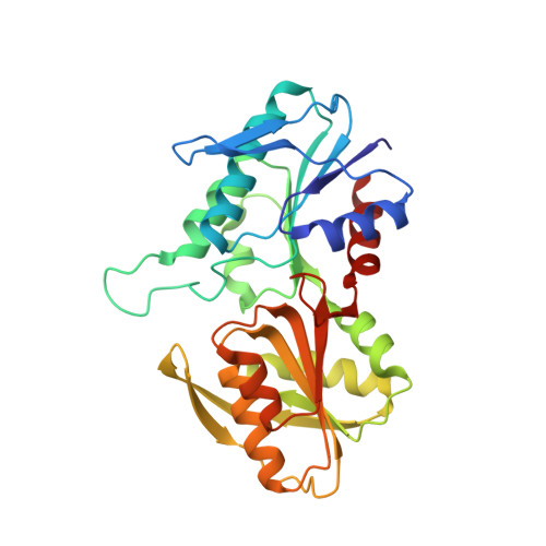
    Phosphoribosyl pyrophosphate (PRPP) synthetase catalyzes the transfer of the pyrophosphate group from ATP to ribose-5-phosphate (R5P) yielding PRPP and AMP. PRPP is an essential metabolite that plays a central role in cellular metabolism. The enzyme from a thermophilic archaeon Thermoplasma volcanium (Tv) was expressed in Escherichia coli, crystallized, and its X-ray molecular structure was determined in a complex with its substrate R5P and with substrate analogs β,γ-methylene ATP and ADP in two monoclinic crystal forms, P2(1). The β,γ-methylene ATP- and the ADP-bound binary structures were determined from crystals grown from ammonium sulfate solutions; these crystals diffracted to 1.8 Å and 1.5 Å resolutions, respectively. Crystals of the ternary complex with ADP-Mg(2+) and R5P were grown from a polyethylene glycol solution in the absence of sulfate ions, and they diffracted to 1.8 Å resolution; the unit cell is approximately double the size of the unit cell of the crystals grown in the presence of sulfate. The Tv PRPP synthetase adopts two conformations, open and closed, at different stages in the catalytic cycle. The binding of substrates, R5P and ATP, occurs with PRPP synthetase in the open conformation, whereas catalysis presumably takes place with PRPP synthetase in the closed conformation. The Tv PRPP synthetase forms a biological dimer in contrast to the tetrameric or hexameric quaternary structures of the Methanocaldococcus jannaschii and Bacillus subtilis PRPP synthetases, respectively.


  • Organizational Affiliation

    Department of Biochemistry, School of Molecular and Systems Medicine, University of Alberta, Edmonton, Alberta, Canada T6G 2H7.


Macromolecules
Find similar proteins by:  (by identity cutoff)  |  3D Structure
Entity ID: 1
MoleculeChains Sequence LengthOrganismDetailsImage
Ribose-phosphate pyrophosphokinase
A, B, C, D
287Thermoplasma volcaniumMutation(s): 0 
Gene Names: prsTV0197TVG0201915TVN0197
EC: 2.7.6.1
UniProt
Find proteins for Q97CA5 (Thermoplasma volcanium (strain ATCC 51530 / DSM 4299 / JCM 9571 / NBRC 15438 / GSS1))
Explore Q97CA5 
Go to UniProtKB:  Q97CA5
Entity Groups  
Sequence Clusters30% Identity50% Identity70% Identity90% Identity95% Identity100% Identity
UniProt GroupQ97CA5
Sequence Annotations
Expand
  • Reference Sequence
Small Molecules
Ligands 4 Unique
IDChains Name / Formula / InChI Key2D Diagram3D Interactions
ADP
Query on ADP

Download Ideal Coordinates CCD File 
G [auth A],
J [auth B],
K [auth C],
P [auth D]
ADENOSINE-5'-DIPHOSPHATE
C10 H15 N5 O10 P2
XTWYTFMLZFPYCI-KQYNXXCUSA-N
HSX
Query on HSX

Download Ideal Coordinates CCD File 
N [auth D]5-O-phosphono-alpha-D-ribofuranose
C5 H11 O8 P
KTVPXOYAKDPRHY-AIHAYLRMSA-N
PO4
Query on PO4

Download Ideal Coordinates CCD File 
E [auth A],
H [auth B],
L [auth C]
PHOSPHATE ION
O4 P
NBIIXXVUZAFLBC-UHFFFAOYSA-K
MG
Query on MG

Download Ideal Coordinates CCD File 
F [auth A],
I [auth B],
M [auth C],
O [auth D]
MAGNESIUM ION
Mg
JLVVSXFLKOJNIY-UHFFFAOYSA-N
Experimental Data & Validation

Experimental Data

  • Method: X-RAY DIFFRACTION
  • Resolution: 1.80 Å
  • R-Value Free: 0.219 
  • R-Value Work: 0.176 
  • R-Value Observed: 0.178 
  • Space Group: P 1 21 1
Unit Cell:
Length ( Å )Angle ( ˚ )
a = 76.599α = 90
b = 61.841β = 103.94
c = 127.938γ = 90
Software Package:
Software NamePurpose
PHENIXmodel building
PHENIXrefinement
HKL-2000data reduction
HKL-2000data scaling
PHENIXphasing

Structure Validation

View Full Validation Report


Ligand Structure Quality Assessment 


Entry History 

Deposition Data

Revision History  (Full details and data files)

  • Version 1.0: 2011-04-20
    Type: Initial release
  • Version 1.1: 2011-07-13
    Changes: Version format compliance
  • Version 1.2: 2012-04-25
    Changes: Database references
  • Version 2.0: 2020-07-29
    Type: Remediation
    Reason: Carbohydrate remediation
    Changes: Atomic model, Data collection, Database references, Derived calculations
  • Version 2.1: 2023-09-06
    Changes: Data collection, Database references, Refinement description, Structure summary