3K8D

Crystal structure of E. coli lipopolysaccharide specific CMP-KDO synthetase in complex with CTP and 2-deoxy-Kdo


Experimental Data Snapshot

  • Method: X-RAY DIFFRACTION
  • Resolution: 1.90 Å
  • R-Value Free: 0.182 
  • R-Value Work: 0.151 
  • R-Value Observed: 0.153 

wwPDB Validation   3D Report Full Report


Ligand Structure Quality Assessment 


This is version 1.4 of the entry. See complete history


Literature

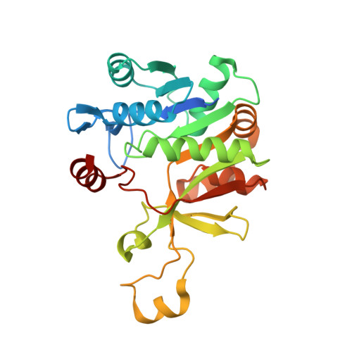
Structure-based mechanism of CMP-2-keto-3-deoxymanno-octulonic acid synthetase: convergent evolution of a sugar-activating enzyme with DNA/RNA polymerases

Heyes, D.J.Levy, C.Lafite, P.Roberts, I.S.Goldrick, M.Stachulski, A.V.Rossington, S.B.Stanford, D.Rigby, S.E.J.Scrutton, N.S.Leys, D.

(2009) J Biol Chem 284: 35514-35523

  • DOI: https://doi.org/10.1074/jbc.M109.056630
  • Primary Citation of Related Structures:  
    3K8D, 3K8E

  • PubMed Abstract: 

    The enzyme CMP-Kdo synthetase (KdsB) catalyzes the addition of 2-keto-3-deoxymanno-octulonic acid (Kdo) to CTP to form CMP-Kdo, a key reaction in the biosynthesis of lipopolysaccharide. The reaction catalyzed by KdsB and the related CMP-acylneuraminate synthase is unique among the sugar-activating enzymes in that the respective sugars are directly coupled to a cytosine monophosphate. Using inhibition studies, in combination with isothermal calorimetry, we show the substrate analogue 2beta-deoxy-Kdo to be a potent competitive inhibitor. The ligand-free Escherichia coli KdsB and ternary complex KdsB-CTP-2beta-deoxy-Kdo crystal structures reveal that Kdo binding leads to active site closure and repositioning of the CTP phosphates and associated Mg(2+) ion (Mg-B). Both ligands occupy conformations compatible with an S(n)2-type attack on the alpha-phosphate by the Kdo 2-hydroxyl group. Based on strong similarity with DNA/RNA polymerases, both in terms of overall chemistry catalyzed as well as active site configuration, we postulate a second Mg(2+) ion (Mg-A) is bound by the catalytically competent KdsB-CTP-Kdo ternary complex. Modeling of this complex reveals the Mg-A coordinated to the conserved Asp(100) and Asp(235) in addition to the CTP alpha-phosphate and both the Kdo carboxylic and 2-hydroxyl groups. EPR measurements on the Mn(2+)-substituted ternary complex support this model. We propose the KdsB/CNS sugar-activating enzymes catalyze the formation of activated sugars, such as the abundant CMP-5-N-acetylneuraminic acid, by recruitment of two Mg(2+) to the active site. Although each metal ion assists in correct positioning of the substrates and activation of the alpha-phosphate, Mg-A is responsible for activation of the sugar-hydroxyl group.


  • Organizational Affiliation

    Manchester Interdisciplinary Biocentre, Faculty of Life Sciences, University of Manchester, 131 Princess Street, Manchester M1 7DN.


Macromolecules
Find similar proteins by:  (by identity cutoff)  |  3D Structure
Entity ID: 1
MoleculeChains Sequence LengthOrganismDetailsImage
3-deoxy-manno-octulosonate cytidylyltransferase
A, B, C, D
264Escherichia coliMutation(s): 0 
EC: 2.7.7.38
UniProt
Find proteins for P04951 (Escherichia coli (strain K12))
Explore P04951 
Go to UniProtKB:  P04951
Entity Groups  
Sequence Clusters30% Identity50% Identity70% Identity90% Identity95% Identity100% Identity
UniProt GroupP04951
Sequence Annotations
Expand
  • Reference Sequence
Small Molecules
Ligands 3 Unique
IDChains Name / Formula / InChI Key2D Diagram3D Interactions
CTP
Query on CTP

Download Ideal Coordinates CCD File 
G [auth A],
J [auth B],
M [auth C],
P [auth D]
CYTIDINE-5'-TRIPHOSPHATE
C9 H16 N3 O14 P3
PCDQPRRSZKQHHS-XVFCMESISA-N
KDO
Query on KDO

Download Ideal Coordinates CCD File 
F [auth A],
I [auth B],
L [auth C],
O [auth D]
3-deoxy-alpha-D-manno-oct-2-ulopyranosonic acid
C8 H14 O8
NNLZBVFSCVTSLA-HXUQBWEZSA-N
MG
Query on MG

Download Ideal Coordinates CCD File 
E [auth A],
H [auth B],
K [auth C],
N [auth D]
MAGNESIUM ION
Mg
JLVVSXFLKOJNIY-UHFFFAOYSA-N
Binding Affinity Annotations 
IDSourceBinding Affinity
KDO Binding MOAD:  3K8D Kd: 1700 (nM) from 1 assay(s)
PDBBind:  3K8D Kd: 1700 (nM) from 1 assay(s)
Experimental Data & Validation

Experimental Data

  • Method: X-RAY DIFFRACTION
  • Resolution: 1.90 Å
  • R-Value Free: 0.182 
  • R-Value Work: 0.151 
  • R-Value Observed: 0.153 
  • Space Group: P 31
Unit Cell:
Length ( Å )Angle ( ˚ )
a = 94.77α = 90
b = 94.77β = 90
c = 153.14γ = 120
Software Package:
Software NamePurpose
DNAdata collection
PHASERphasing
PHENIXrefinement
XDSdata reduction
XSCALEdata scaling

Structure Validation

View Full Validation Report


Ligand Structure Quality Assessment 


Entry History 

Deposition Data

Revision History  (Full details and data files)

  • Version 1.0: 2009-11-10
    Type: Initial release
  • Version 1.1: 2011-07-13
    Changes: Version format compliance
  • Version 1.2: 2012-12-05
    Changes: Database references
  • Version 1.3: 2020-07-29
    Type: Remediation
    Reason: Carbohydrate remediation
    Changes: Advisory, Data collection, Database references, Derived calculations, Structure summary
  • Version 1.4: 2023-11-01
    Changes: Data collection, Database references, Refinement description, Structure summary