3K6N

Crystal structure of the S225E mutant Kir3.1 cytoplasmic pore domain


Experimental Data Snapshot

  • Method: X-RAY DIFFRACTION
  • Resolution: 2.00 Å
  • R-Value Free: 0.266 
  • R-Value Work: 0.254 

wwPDB Validation   3D Report Full Report


This is version 1.5 of the entry. See complete history


Literature

Physical determinants of strong voltage sensitivity of K(+) channel block.

Xu, Y.Shin, H.G.Szep, S.Lu, Z.

(2009) Nat Struct Mol Biol 16: 1252-1258

  • DOI: https://doi.org/10.1038/nsmb.1717
  • Primary Citation of Related Structures:  
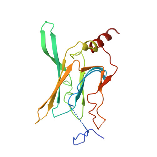
    3K6N

  • PubMed Abstract: 

    Strong voltage sensitivity of inward-rectifier K(+) (Kir) channels has been hypothesized to arise primarily from an intracellular blocker displacing up to five K(+) ions from the wide, intracellular part of the ion conduction pore outwardly across the narrow ion-selectivity filter. The validity of this hypothesis depends on two assumptions: (i) that five ion sites are located intracellular to the filter and (ii) that the blocker can force essentially unidirectional K(+) movement in a pore region generally wider than the combined dimensions of the blocker plus a K(+) ion. Here we present a crystal structure of the cytoplasmic portion of a Kir channel with five ions bound and demonstrate that a constriction near the intracellular end of the pore, acting as a gasket, prevents K(+) ions from bypassing the blocker. This heretofore unrecognized 'gasket' ensures that the blocker can effectively displace K(+) ions across the selectivity filter to generate exceedingly strong voltage sensitivity.


  • Organizational Affiliation

    Department of Physiology, Howard Hughes Medical Institute, University of Pennsylvania, Philadelphia, Pennsylvania, USA.


Macromolecules
Find similar proteins by:  (by identity cutoff)  |  3D Structure
Entity ID: 1
MoleculeChains Sequence LengthOrganismDetailsImage
G protein-activated inward rectifier potassium channel 1207Mus musculusMutation(s): 1 
Gene Names: Girk1Kcnj3
Membrane Entity: Yes 
UniProt & NIH Common Fund Data Resources
Find proteins for P63250 (Mus musculus)
Explore P63250 
Go to UniProtKB:  P63250
IMPC:  MGI:104742
Entity Groups  
Sequence Clusters30% Identity50% Identity70% Identity90% Identity95% Identity100% Identity
UniProt GroupP63250
Sequence Annotations
Expand
  • Reference Sequence
Experimental Data & Validation

Experimental Data

  • Method: X-RAY DIFFRACTION
  • Resolution: 2.00 Å
  • R-Value Free: 0.266 
  • R-Value Work: 0.254 
  • Space Group: P 4 21 2
Unit Cell:
Length ( Å )Angle ( ˚ )
a = 77.455α = 90
b = 77.455β = 90
c = 86.723γ = 90
Software Package:
Software NamePurpose
DENZOdata reduction
SCALEPACKdata scaling
AMoREphasing
CNSrefinement
PDB_EXTRACTdata extraction
HKL-2000data collection
HKL-2000data reduction
HKL-2000data scaling

Structure Validation

View Full Validation Report


Entry History 

Deposition Data

Revision History  (Full details and data files)

  • Version 1.0: 2009-11-17
    Type: Initial release
  • Version 1.1: 2011-07-13
    Changes: Version format compliance
  • Version 1.2: 2017-08-02
    Changes: Refinement description, Source and taxonomy
  • Version 1.3: 2017-11-01
    Changes: Refinement description
  • Version 1.4: 2021-10-13
    Changes: Database references, Derived calculations
  • Version 1.5: 2023-09-06
    Changes: Data collection, Refinement description