3IR8

Red fluorescent protein mKeima at pH 7.0


Experimental Data Snapshot

  • Method: X-RAY DIFFRACTION
  • Resolution: 1.63 Å
  • R-Value Free: 0.235 
  • R-Value Work: 0.193 
  • R-Value Observed: 0.195 

wwPDB Validation   3D Report Full Report


This is version 2.0 of the entry. See complete history


Literature

Excited state proton transfer in the red fluorescent protein mKeima.

Henderson, J.N.Osborn, M.F.Koon, N.Gepshtein, R.Huppert, D.Remington, S.J.

(2009) J Am Chem Soc 131: 13212-13213

  • DOI: https://doi.org/10.1021/ja904665x
  • Primary Citation of Related Structures:  
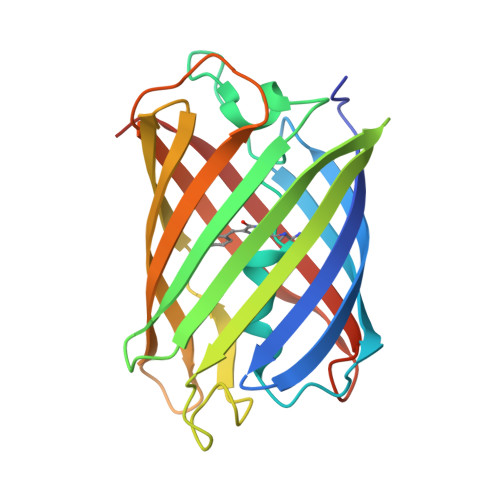
    3IR8

  • PubMed Abstract: 

    mKeima is an unusual monomeric red fluorescent protein (lambda(em)(max) approximately 620 nm) that is maximally excited in the blue (lambda(ex)(max) approximately 440 nm). The large Stokes shift suggests that the chromophore is normally protonated. A 1.63 A resolution structure of mKeima reveals the chromophore to be imbedded in a novel hydrogen bond network, different than in GFP, which could support proton transfer from the chromophore hydroxyl, via Ser142, to Asp157. At low temperatures the emission contains a green component (lambda(em)(max) approximately 535 nm), enhanced by deuterium substitution, presumably resulting from reduced proton transfer efficiency. Ultrafast pump/probe studies reveal a rising component in the 610 nm emission with a lifetime of approximately 4 ps, characterizing the rate of proton transfer. Mutation of Asp157 to neutral Asn changes the chromophore resting charge state to anionic (lambda(ex)(max) approximately 565 nm, lambda(em)(max) approximately 620 nm). Thus, excited state proton transfer (ESPT) explains the large Stokes shift. This work unambiguously characterizes green emission from the protonated acylimine chromophore of red fluorescent proteins.


  • Organizational Affiliation

    Institute of Molecular Biology and Department of Physics, University of Oregon, Eugene, Oregon 97403-1229, USA.


Macromolecules
Find similar proteins by:  (by identity cutoff)  |  3D Structure
Entity ID: 1
MoleculeChains Sequence LengthOrganismDetailsImage
Large stokes shift fluorescent protein
A, B
221Montipora sp. 20Mutation(s): 1 
Gene Names: m-keima
UniProt
Find proteins for Q1JV70 (Montipora sp. 20)
Explore Q1JV70 
Go to UniProtKB:  Q1JV70
Entity Groups  
Sequence Clusters30% Identity50% Identity70% Identity90% Identity95% Identity100% Identity
UniProt GroupQ1JV70
Sequence Annotations
Expand
  • Reference Sequence
Small Molecules
Modified Residues  1 Unique
IDChains TypeFormula2D DiagramParent
CRQ
Query on CRQ
A, B
L-PEPTIDE LINKINGC16 H16 N4 O5GLN, TYR, GLY
Experimental Data & Validation

Experimental Data

  • Method: X-RAY DIFFRACTION
  • Resolution: 1.63 Å
  • R-Value Free: 0.235 
  • R-Value Work: 0.193 
  • R-Value Observed: 0.195 
  • Space Group: P 21 21 21
Unit Cell:
Length ( Å )Angle ( ˚ )
a = 66.959α = 90
b = 75.686β = 90
c = 85.674γ = 90
Software Package:
Software NamePurpose
DENZOdata reduction
SCALEPACKdata scaling
EPMRphasing
REFMACrefinement
PDB_EXTRACTdata extraction
HKL-2000data collection

Structure Validation

View Full Validation Report


Entry History 

Deposition Data

Revision History  (Full details and data files)

  • Version 1.0: 2009-09-08
    Type: Initial release
  • Version 1.1: 2011-07-13
    Changes: Version format compliance
  • Version 1.2: 2017-11-01
    Changes: Refinement description
  • Version 1.3: 2023-09-06
    Changes: Data collection, Database references, Derived calculations, Refinement description
  • Version 2.0: 2023-11-15
    Changes: Atomic model, Data collection, Derived calculations