


Crystal structure of the human MOCS3 rhodanese-like domain
Bacik, J.P., Walker, J.R., Lopez, L., Li, Y., Weigelt, J., Bountra, C., Arrowsmith, C.H., Edwards, A.M., Bochkarev, A., Dhe-Paganon, S.To be published.
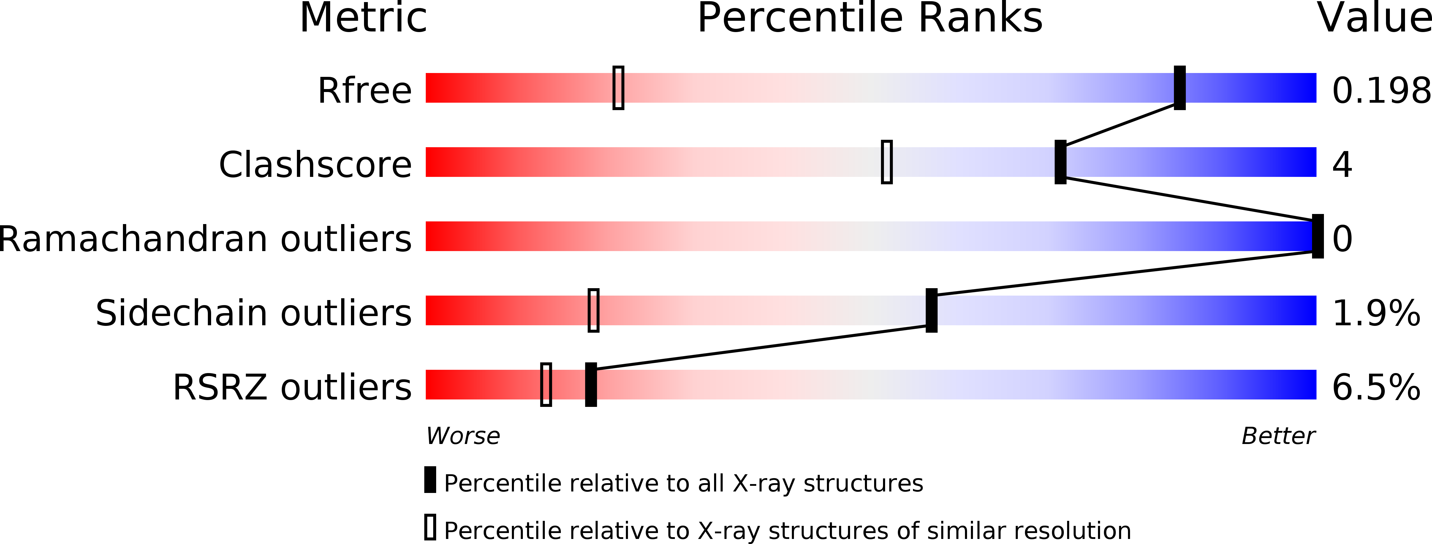
Experimental Data Snapshot
wwPDB Validation 3D Report Full Report
Entity ID: 1 | |||||
|---|---|---|---|---|---|
| Molecule | Chains | Sequence Length | Organism | Details | Image |
| Adenylyltransferase and sulfurtransferase MOCS3 | 127 | Homo sapiens | Mutation(s): 0 Gene Names: MOCS3, MOCS3_HUMAN, UBA4 EC: 2.7.7 (PDB Primary Data), 2.8.1 (PDB Primary Data) |  | |
UniProt & NIH Common Fund Data Resources | |||||
Find proteins for O95396 (Homo sapiens) Explore O95396 Go to UniProtKB: O95396 | |||||
PHAROS: O95396 GTEx: ENSG00000124217 | |||||
Entity Groups | |||||
| Sequence Clusters | 30% Identity50% Identity70% Identity90% Identity95% Identity100% Identity | ||||
| UniProt Group | O95396 | ||||
Sequence AnnotationsExpand | |||||
| |||||
| Modified Residues 1 Unique | |||||
|---|---|---|---|---|---|
| ID | Chains | Type | Formula | 2D Diagram | Parent |
| MSE Query on MSE | A | L-PEPTIDE LINKING | C5 H11 N O2 Se |  | MET |
| Length ( Å ) | Angle ( ˚ ) |
|---|---|
| a = 36.817 | α = 90 |
| b = 31.911 | β = 110.16 |
| c = 50.64 | γ = 90 |
| Software Name | Purpose |
|---|---|
| HKL-2000 | data collection |
| PHENIX | model building |
| PHENIX | refinement |
| HKL-2000 | data reduction |
| HKL-2000 | data scaling |
| PHENIX | phasing |
RCSB PDB (citation) is hosted by
RCSB PDB is a member of the

