Substrate-mediated Stabilization of a Tetrameric Drug Target Reveals Achilles Heel in Anthrax.
Voss, J.E., Scally, S.W., Taylor, N.L., Atkinson, S.C., Griffin, M.D., Hutton, C.A., Parker, M.W., Alderton, M.R., Gerrard, J.A., Dobson, R.C., Dogovski, C., Perugini, M.A.(2010) J Biol Chem 285: 5188-5195
- PubMed: 19948665
- DOI: https://doi.org/10.1074/jbc.M109.038166
- Primary Citation of Related Structures:
3HIJ - PubMed Abstract:
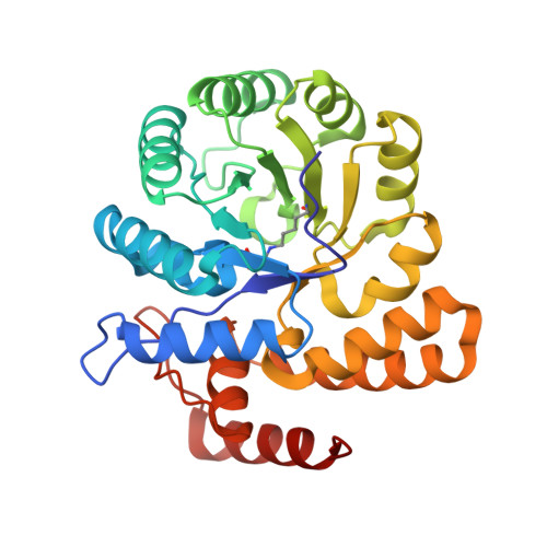
Bacillus anthracis is a gram-positive spore-forming bacterium that causes anthrax. With the increased threat of anthrax in biowarfare, there is an urgent need to characterize new antimicrobial targets from B. anthracis. One such target is dihydrodipicolinate synthase (DHDPS), which catalyzes the committed step in the pathway yielding meso-diaminopimelate and lysine. In this study, we employed CD spectroscopy to demonstrate that the thermostability of DHDPS from B. anthracis (Ba-DHDPS) is significantly enhanced in the presence of the substrate, pyruvate. Analytical ultracentrifugation studies show that the tetramer-dimer dissociation constant of the enzyme is 3-fold tighter in the presence of pyruvate compared with the apo form. To examine the significance of this substrate-mediated stabilization phenomenon, a dimeric mutant of Ba-DHDPS (L170E/G191E) was generated and shown to have markedly reduced activity compared with the wild-type tetramer. This demonstrates that the substrate, pyruvate, stabilizes the active form of the enzyme. We next determined the high resolution (2.15 A) crystal structure of Ba-DHDPS in complex with pyruvate (3HIJ) and compared this to the apo structure (1XL9). Structural analyses show that there is a significant (91 A(2)) increase in buried surface area at the tetramerization interface of the pyruvate-bound structure. This study describes a new mechanism for stabilization of the active oligomeric form of an antibiotic target from B. anthracis and reveals an "Achilles heel" that can be exploited in structure-based drug design.
Organizational Affiliation:
Department of Biochemistry and Molecular Biology, University of Melbourne, Parkville, Victoria 3010, Australia.