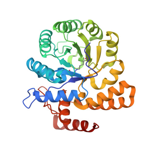
3HIJ

Crystal structure of dihydrodipicolinate synthase from Bacillus anthracis in complex with its substrate, pyruvate


Experimental Data Snapshot

  • Method: X-RAY DIFFRACTION
  • Resolution: 2.15 Å
  • R-Value Free: 0.210 
  • R-Value Work: 0.153 
  • R-Value Observed: 0.155 

wwPDB Validation   3D Report Full Report


This is version 1.1 of the entry. See complete history


Literature

Substrate-mediated Stabilization of a Tetrameric Drug Target Reveals Achilles Heel in Anthrax.

Voss, J.E.Scally, S.W.Taylor, N.L.Atkinson, S.C.Griffin, M.D.Hutton, C.A.Parker, M.W.Alderton, M.R.Gerrard, J.A.Dobson, R.C.Dogovski, C.Perugini, M.A.

(2010) J Biol Chem 285: 5188-5195

  • DOI: https://doi.org/10.1074/jbc.M109.038166
  • Primary Citation of Related Structures:  
    3HIJ

  • PubMed Abstract: 

    Bacillus anthracis is a gram-positive spore-forming bacterium that causes anthrax. With the increased threat of anthrax in biowarfare, there is an urgent need to characterize new antimicrobial targets from B. anthracis. One such target is dihydrodipicolinate synthase (DHDPS), which catalyzes the committed step in the pathway yielding meso-diaminopimelate and lysine. In this study, we employed CD spectroscopy to demonstrate that the thermostability of DHDPS from B. anthracis (Ba-DHDPS) is significantly enhanced in the presence of the substrate, pyruvate. Analytical ultracentrifugation studies show that the tetramer-dimer dissociation constant of the enzyme is 3-fold tighter in the presence of pyruvate compared with the apo form. To examine the significance of this substrate-mediated stabilization phenomenon, a dimeric mutant of Ba-DHDPS (L170E/G191E) was generated and shown to have markedly reduced activity compared with the wild-type tetramer. This demonstrates that the substrate, pyruvate, stabilizes the active form of the enzyme. We next determined the high resolution (2.15 A) crystal structure of Ba-DHDPS in complex with pyruvate (3HIJ) and compared this to the apo structure (1XL9). Structural analyses show that there is a significant (91 A(2)) increase in buried surface area at the tetramerization interface of the pyruvate-bound structure. This study describes a new mechanism for stabilization of the active oligomeric form of an antibiotic target from B. anthracis and reveals an "Achilles heel" that can be exploited in structure-based drug design.


  • Organizational Affiliation

    Department of Biochemistry and Molecular Biology, University of Melbourne, Parkville, Victoria 3010, Australia.


Macromolecules
Find similar proteins by:  (by identity cutoff)  |  3D Structure
Entity ID: 1
MoleculeChains Sequence LengthOrganismDetailsImage
Dihydrodipicolinate synthase
A, B, C, D
292Bacillus anthracisMutation(s): 0 
Gene Names: dapA-2dapA2BAS3650BA_3935GBAA3935GBAA_3935
EC: 4.3.3.7
UniProt
Find proteins for A0A6L7H340 (Bacillus anthracis)
Explore A0A6L7H340 
Go to UniProtKB:  A0A6L7H340
Entity Groups  
Sequence Clusters30% Identity50% Identity70% Identity90% Identity95% Identity100% Identity
UniProt GroupA0A6L7H340
Sequence Annotations
Expand
  • Reference Sequence
Small Molecules
Ligands 2 Unique
IDChains Name / Formula / InChI Key2D Diagram3D Interactions
GOL
Query on GOL

Download Ideal Coordinates CCD File 
E [auth A],
G [auth B],
H [auth B],
K [auth D]
GLYCEROL
C3 H8 O3
PEDCQBHIVMGVHV-UHFFFAOYSA-N
NA
Query on NA

Download Ideal Coordinates CCD File 
F [auth A],
I [auth B],
J [auth C],
L [auth D]
SODIUM ION
Na
FKNQFGJONOIPTF-UHFFFAOYSA-N
Modified Residues  1 Unique
IDChains TypeFormula2D DiagramParent
KPI
Query on KPI
A, B, C, D
L-PEPTIDE LINKINGC9 H16 N2 O4LYS
Experimental Data & Validation

Experimental Data

  • Method: X-RAY DIFFRACTION
  • Resolution: 2.15 Å
  • R-Value Free: 0.210 
  • R-Value Work: 0.153 
  • R-Value Observed: 0.155 
  • Space Group: P 21 21 21
Unit Cell:
Length ( Å )Angle ( ˚ )
a = 84.484α = 90
b = 124.617β = 90
c = 130.979γ = 90
Software Package:
Software NamePurpose
MOSFLMdata reduction
PHASERphasing
REFMACrefinement
PDB_EXTRACTdata extraction
ADSCdata collection
SCALAdata scaling

Structure Validation

View Full Validation Report


Entry History 

Deposition Data

Revision History  (Full details and data files)

  • Version 1.0: 2009-11-24
    Type: Initial release
  • Version 1.1: 2011-07-13
    Changes: Non-polymer description, Version format compliance