3GX5

Crystal structure of T. tencongensis SAM-I riboswitch variant A94G/U34 bound with SAM


Experimental Data Snapshot

  • Method: X-RAY DIFFRACTION
  • Resolution: 2.40 Å
  • R-Value Free: 0.263 
  • R-Value Work: 0.202 
  • R-Value Observed: 0.208 

wwPDB Validation   3D Report Full Report


Ligand Structure Quality Assessment 


This is version 1.2 of the entry. See complete history


Literature

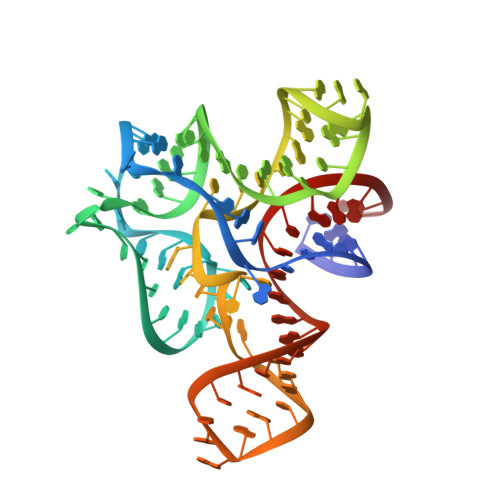
Discrimination between Closely Related Cellular Metabolites by the SAM-I Riboswitch.

Montange, R.K.Mondragon, E.van Tyne, D.Garst, A.D.Ceres, P.Batey, R.T.

(2010) J Mol Biol 396: 761-772

  • DOI: https://doi.org/10.1016/j.jmb.2009.12.007
  • Primary Citation of Related Structures:  
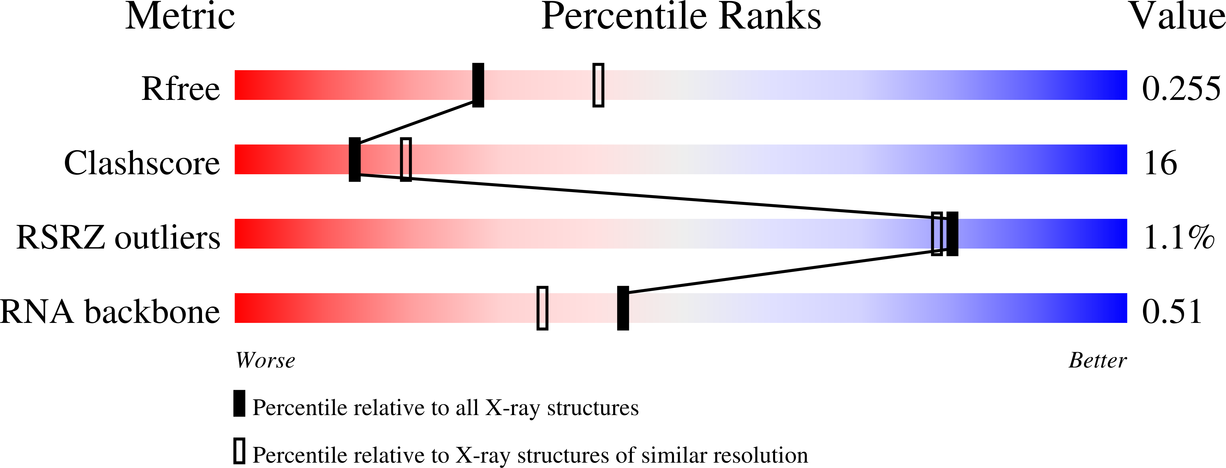
    3GX2, 3GX3, 3GX5, 3GX6, 3GX7

  • PubMed Abstract: 

    The SAM-I riboswitch is a cis-acting element of genetic control found in bacterial mRNAs that specifically binds S-adenosylmethionine (SAM). We previously determined the 2.9-A X-ray crystal structure of the effector-binding domain of this RNA element, revealing details of RNA-ligand recognition. To improve this structure, variations were made to the RNA sequence to alter lattice contacts, resulting in a 0.5-A improvement in crystallographic resolution and allowing for a more accurate refinement of the crystallographic model. The basis for SAM specificity was addressed by a structural analysis of the RNA complexed to S-adenosylhomocysteine (SAH) and sinefungin and by measuring the affinity of SAM and SAH for a series of mutants using isothermal titration calorimetry. These data illustrate the importance of two universally conserved base pairs in the RNA that form electrostatic interactions with the positively charged sulfonium group of SAM, thereby providing a basis for discrimination between SAM and SAH.


  • Organizational Affiliation

    Department of Chemistry and Biochemistry, University of Colorado, Boulder, Campus Box 215, Boulder, CO 80309-0215, USA.


Macromolecules
Find similar nucleic acids by:  (by identity cutoff)  |  3D Structure
Entity ID: 1
MoleculeChains LengthOrganismImage
RNA (94-MER)94N/A
Sequence Annotations
Expand
  • Reference Sequence
Small Molecules
Ligands 3 Unique
IDChains Name / Formula / InChI Key2D Diagram3D Interactions
SAM
Query on SAM

Download Ideal Coordinates CCD File 
B [auth A]S-ADENOSYLMETHIONINE
C15 H22 N6 O5 S
MEFKEPWMEQBLKI-FCKMPRQPSA-N
K
Query on K

Download Ideal Coordinates CCD File 
K [auth A]POTASSIUM ION
K
NPYPAHLBTDXSSS-UHFFFAOYSA-N
MG
Query on MG

Download Ideal Coordinates CCD File 
C [auth A]
D [auth A]
E [auth A]
F [auth A]
G [auth A]
C [auth A],
D [auth A],
E [auth A],
F [auth A],
G [auth A],
H [auth A],
I [auth A],
J [auth A]
MAGNESIUM ION
Mg
JLVVSXFLKOJNIY-UHFFFAOYSA-N
Experimental Data & Validation

Experimental Data

  • Method: X-RAY DIFFRACTION
  • Resolution: 2.40 Å
  • R-Value Free: 0.263 
  • R-Value Work: 0.202 
  • R-Value Observed: 0.208 
  • Space Group: P 43 21 2
Unit Cell:
Length ( Å )Angle ( ˚ )
a = 62.71α = 90
b = 62.71β = 90
c = 158.68γ = 90
Software Package:
Software NamePurpose
HKL-2000data collection
PHENIXmodel building
PHENIXrefinement
HKL-2000data reduction
HKL-2000data scaling
PHENIXphasing

Structure Validation

View Full Validation Report


Ligand Structure Quality Assessment 


Entry History 

Deposition Data

Revision History  (Full details and data files)

  • Version 1.0: 2010-01-12
    Type: Initial release
  • Version 1.1: 2011-07-13
    Changes: Version format compliance
  • Version 1.2: 2023-09-06
    Changes: Data collection, Database references, Derived calculations, Refinement description