Two Site-Directed Mutations Are Required for the Conversion of a Sugar Dehydratase into an Aminotransferase.
Cook, P.D., Kubiak, R.L., Toomey, D.P., Holden, H.M.(2009) Biochemistry 48: 5246-5253
- PubMed: 19402712
- DOI: https://doi.org/10.1021/bi9005545
- Primary Citation of Related Structures:
3GR9 - PubMed Abstract:
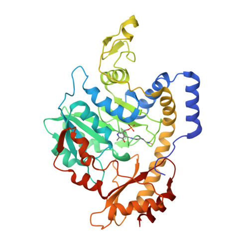
L-colitose and d-perosamine are unusual sugars found in the O-antigens of some Gram-negative bacteria such as Escherichia coli, Vibrio cholerae, and Salmonella enterica, among others. The biosynthetic pathways for these two sugars begin with the formation of GDP-mannose from d-mannose 1-phosphate and GTP followed by the subsequent dehydration and oxidation of GDP-mannose to yield GDP-4-keto-6-deoxymannose. Following the production of GDP-4-keto-6-deoxymannose, the two pathways diverge. In the case of GDP-perosamine biosynthesis, the next step involves an amination reaction at the C-4' position of the sugar, whereas in GDP-colitose production, the 3'-hydroxyl group is removed. The enzymes catalyzing these reactions are GDP-perosamine synthase and GDP-4-keto-6-deoxymannose-3-dehydratase (ColD), respectively. Both of these enzymes are pyridoxal 5'-phosphate (PLP) dependent, and their three-dimensional structures place them into the well-characterized aspartate aminotransferase superfamily. A comparison of the active site architecture of ColD from E. coli (strain 5a, type O55:H7) to that of GDP-perosamine synthase from Caulobacter crescentus CB15 suggested that only two mutations would be required to convert ColD into an aminotransferase. Here we present a combined structural and functional analysis of the ColD S187N/H188K mutant protein that, indeed, has been converted from a sugar dehydratase into an aminotransferase.
Organizational Affiliation:
Department of Biochemistry, University of Wisconsin, Madison, Wisconsin 53706, USA.