3FPU

The crystallographic structure of the Complex between Evasin-1 and CCL3


Experimental Data Snapshot

  • Method: X-RAY DIFFRACTION
  • Resolution: 1.76 Å
  • R-Value Free: 0.285 
  • R-Value Work: 0.231 
  • R-Value Observed: 0.234 

wwPDB Validation   3D Report Full Report


This is version 1.4 of the entry. See complete history


Literature

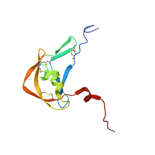
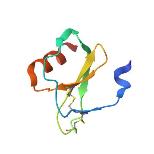
Structural basis of chemokine sequestration by a tick chemokine binding protein: the crystal structure of the complex between Evasin-1 and CCL3

Dias, J.M.Losberger, C.Deruaz, M.Power, C.A.Proudfoot, A.E.I.Shaw, J.P.

(2009) PLoS One 4

  • DOI: https://doi.org/10.1371/journal.pone.0008514
  • Primary Citation of Related Structures:  
    3FPR, 3FPT, 3FPU

  • PubMed Abstract: 

    Chemokines are a subset of cytokines responsible for controlling the cellular migration of inflammatory cells through interaction with seven transmembrane G protein-coupled receptors. The blocking of a chemokine-receptor interaction results in a reduced inflammatory response, and represents a possible anti-inflammatory strategy, a strategy that is already employed by some virus and parasites. Anti-chemokine activity has been described in the extracts of tick salivary glands, and we have recently described the cloning and characterization of such chemokine binding proteins from the salivary glands, which we have named Evasins.


  • Organizational Affiliation

    Merck Serono Geneva Research Center, Merck Serono S.A., Geneva, Switzerland.


Macromolecules
Find similar proteins by:  (by identity cutoff)  |  3D Structure
Entity ID: 1
MoleculeChains Sequence LengthOrganismDetailsImage
Evasin-1100Rhipicephalus sanguineusMutation(s): 0 
UniProt
Find proteins for P0C8E7 (Rhipicephalus sanguineus)
Explore P0C8E7 
Go to UniProtKB:  P0C8E7
Entity Groups  
Sequence Clusters30% Identity50% Identity70% Identity90% Identity95% Identity100% Identity
UniProt GroupP0C8E7
Sequence Annotations
Expand
  • Reference Sequence
Find similar proteins by:  (by identity cutoff)  |  3D Structure
Entity ID: 2
MoleculeChains Sequence LengthOrganismDetailsImage
C-C motif chemokine 370Homo sapiensMutation(s): 1 
UniProt & NIH Common Fund Data Resources
Find proteins for P10147 (Homo sapiens)
Explore P10147 
Go to UniProtKB:  P10147
PHAROS:  P10147
GTEx:  ENSG00000277632 
Entity Groups  
Sequence Clusters30% Identity50% Identity70% Identity90% Identity95% Identity100% Identity
UniProt GroupP10147
Sequence Annotations
Expand
  • Reference Sequence
Experimental Data & Validation

Experimental Data

  • Method: X-RAY DIFFRACTION
  • Resolution: 1.76 Å
  • R-Value Free: 0.285 
  • R-Value Work: 0.231 
  • R-Value Observed: 0.234 
  • Space Group: P 43 3 2
Unit Cell:
Length ( Å )Angle ( ˚ )
a = 104.384α = 90
b = 104.384β = 90
c = 104.384γ = 90
Software Package:
Software NamePurpose
HKL-2000data collection
AMoREphasing
REFMACrefinement
DENZOdata reduction
SCALEPACKdata scaling

Structure Validation

View Full Validation Report


Entry History 

Deposition Data

Revision History  (Full details and data files)

  • Version 1.0: 2010-01-12
    Type: Initial release
  • Version 1.1: 2011-07-13
    Changes: Version format compliance
  • Version 1.2: 2013-10-16
    Changes: Derived calculations
  • Version 1.3: 2021-11-10
    Changes: Database references, Derived calculations
  • Version 1.4: 2023-11-01
    Changes: Data collection, Refinement description