3CX4

Crystal Structure of E.coli GS mutant E377A in complex with ADP and oligosaccharides


Experimental Data Snapshot

  • Method: X-RAY DIFFRACTION
  • Resolution: 2.29 Å
  • R-Value Free: 0.221 
  • R-Value Work: 0.186 
  • R-Value Observed: 0.187 

wwPDB Validation   3D Report Full Report


Ligand Structure Quality Assessment 


This is version 2.2 of the entry. See complete history


Literature

Oligosaccharide binding in Escherichia coli glycogen synthase.

Sheng, F.Yep, A.Feng, L.Preiss, J.Geiger, J.H.

(2009) Biochemistry 48: 10089-10097

  • DOI: https://doi.org/10.1021/bi900916t
  • Primary Citation of Related Structures:  
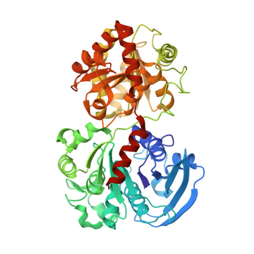
    3CX4

  • PubMed Abstract: 

    Glycogen/starch synthase elongates glucan chains and is the key enzyme in the synthesis of glycogen in bacteria and starch in plants. Cocrystallization of Escherichia coli wild-type glycogen synthase (GS) with substrate ADPGlc and the glucan acceptor mimic HEPPSO produced a closed form of GS and suggests that domain-domain closure accompanies glycogen synthesis. Cocrystallization of the inactive GS mutant E377A with substrate ADPGlc and oligosaccharide results in the first oligosaccharide-bound glycogen synthase structure. Four bound oligosaccharides are observed, one in the interdomain cleft (G6a) and three on the N-terminal domain surface (G6b, G6c, and G6d). Extending from the center of the enzyme to the interdomain cleft opening, G6a mostly interacts with the highly conserved N-terminal domain residues lining the cleft of GS. The surface-bound oligosaccharides G6c and G6d have less interaction with enzyme and exhibit a more curled, helixlike structural arrangement. The observation that oligosaccharides bind only to the N-terminal domain of GS suggests that glycogen in vivo probably binds to only one side of the enzyme to ensure unencumbered interdomain movement, which is required for efficient, continuous glucan-chain synthesis.


  • Organizational Affiliation

    Department of Chemistry, Michigan State University, East Lansing, Michigan 48824, USA.


Macromolecules
Find similar proteins by:  (by identity cutoff)  |  3D Structure
Entity ID: 1
MoleculeChains Sequence LengthOrganismDetailsImage
Glycogen synthase485Escherichia coliMutation(s): 1 
Gene Names: glgA
EC: 2.4.1.21
UniProt
Find proteins for P0A6U8 (Escherichia coli (strain K12))
Explore P0A6U8 
Go to UniProtKB:  P0A6U8
Entity Groups  
Sequence Clusters30% Identity50% Identity70% Identity90% Identity95% Identity100% Identity
UniProt GroupP0A6U8
Sequence Annotations
Expand
  • Reference Sequence
Oligosaccharides

Help

Entity ID: 2
MoleculeChains Length2D Diagram Glycosylation3D Interactions
alpha-D-glucopyranose-(1-4)-alpha-D-glucopyranose-(1-4)-alpha-D-glucopyranose
B
3N/A
Glycosylation Resources
GlyTouCan:  G96370VA
GlyCosmos:  G96370VA
GlyGen:  G96370VA
Entity ID: 3
MoleculeChains Length2D Diagram Glycosylation3D Interactions
alpha-D-glucopyranose-(1-4)-alpha-D-glucopyranose
C
2N/A
Glycosylation Resources
GlyTouCan:  G07411ON
GlyCosmos:  G07411ON
Entity ID: 4
MoleculeChains Length2D Diagram Glycosylation3D Interactions
alpha-D-glucopyranose-(1-4)-alpha-D-glucopyranose-(1-4)-alpha-D-glucopyranose-(1-4)-alpha-D-glucopyranose-(1-4)-alpha-D-glucopyranose
D
5N/A
Glycosylation Resources
GlyTouCan:  G50146AM
GlyCosmos:  G50146AM
Entity ID: 5
MoleculeChains Length2D Diagram Glycosylation3D Interactions
alpha-D-glucopyranose-(1-4)-alpha-D-glucopyranose-(1-4)-alpha-D-glucopyranose-(1-4)-alpha-D-glucopyranose-(1-4)-alpha-D-glucopyranose-(1-4)-beta-D-glucopyranose
E
6N/A
Glycosylation Resources
GlyTouCan:  G86211GT
GlyCosmos:  G86211GT
GlyGen:  G86211GT
Small Molecules
Ligands 4 Unique
IDChains Name / Formula / InChI Key2D Diagram3D Interactions
ADP
Query on ADP

Download Ideal Coordinates CCD File 
H [auth A]ADENOSINE-5'-DIPHOSPHATE
C10 H15 N5 O10 P2
XTWYTFMLZFPYCI-KQYNXXCUSA-N
250
Query on 250

Download Ideal Coordinates CCD File 
I [auth A](2R)-2-hydroxy-3-[4-(2-hydroxyethyl)piperazin-1-yl]propane-1-sulfonic acid
C9 H20 N2 O5 S
GIZQLVPDAOBAFN-SECBINFHSA-N
ETE
Query on ETE

Download Ideal Coordinates CCD File 
J [auth A]
K [auth A]
L [auth A]
M [auth A]
N [auth A]
J [auth A],
K [auth A],
L [auth A],
M [auth A],
N [auth A],
O [auth A],
P [auth A],
Q [auth A],
R [auth A],
S [auth A],
T [auth A],
U [auth A]
2-{2-[2-2-(METHOXY-ETHOXY)-ETHOXY]-ETHOXY}-ETHANOL
C9 H20 O5
ZNYRFEPBTVGZDN-UHFFFAOYSA-N
NA
Query on NA

Download Ideal Coordinates CCD File 
F [auth A],
G [auth A]
SODIUM ION
Na
FKNQFGJONOIPTF-UHFFFAOYSA-N
Experimental Data & Validation

Experimental Data

  • Method: X-RAY DIFFRACTION
  • Resolution: 2.29 Å
  • R-Value Free: 0.221 
  • R-Value Work: 0.186 
  • R-Value Observed: 0.187 
  • Space Group: I 41
Unit Cell:
Length ( Å )Angle ( ˚ )
a = 126.324α = 90
b = 126.324β = 90
c = 152.334γ = 90
Software Package:
Software NamePurpose
MOLREPphasing
REFMACrefinement
PDB_EXTRACTdata extraction
HKL-2000data collection
X-GENdata reduction

Structure Validation

View Full Validation Report


Ligand Structure Quality Assessment 


Entry History 

Deposition Data

Revision History  (Full details and data files)

  • Version 1.0: 2009-04-28
    Type: Initial release
  • Version 1.1: 2011-07-13
    Changes: Version format compliance
  • Version 2.0: 2020-07-29
    Type: Remediation
    Reason: Carbohydrate remediation
    Changes: Advisory, Atomic model, Data collection, Database references, Derived calculations, Structure summary
  • Version 2.1: 2021-10-20
    Changes: Advisory, Database references, Structure summary
  • Version 2.2: 2023-08-30
    Changes: Data collection, Refinement description