3ACL

Crystal Structure of Human Pirin in complex with Triphenyl Compound


Experimental Data Snapshot

  • Method: X-RAY DIFFRACTION
  • Resolution: 2.35 Å
  • R-Value Free: 0.246 
  • R-Value Work: 0.203 
  • R-Value Observed: 0.203 

wwPDB Validation   3D Report Full Report


Ligand Structure Quality Assessment 


This is version 1.2 of the entry. See complete history


Literature

A small-molecule inhibitor shows that pirin regulates migration of melanoma cells

Miyazaki, I.Simizu, S.Okumura, H.Takagi, S.Osada, H.

(2010) Nat Chem Biol 6: 667-673

  • DOI: https://doi.org/10.1038/nchembio.423
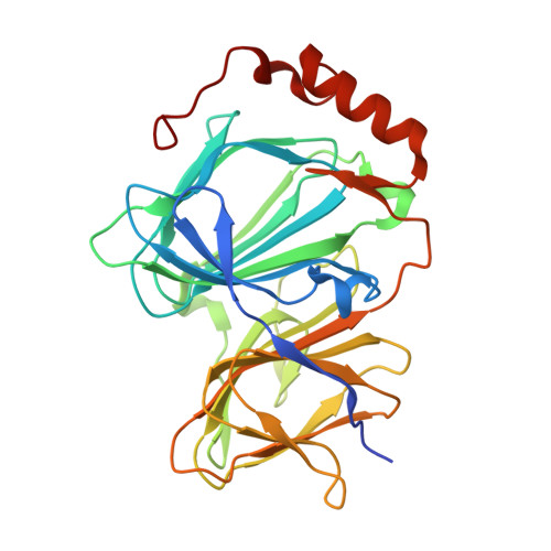
  • Primary Citation of Related Structures:  
    3ACL

  • PubMed Abstract: 

    The discovery of small molecules that bind to a specific target and disrupt the function of proteins is an important step in chemical biology, especially for poorly characterized proteins. Human pirin is a nuclear protein of unknown function that is widely expressed in punctate subnuclear structures in human tissues. Here, we report the discovery of a small molecule that binds to pirin. We determined how the small molecule bound to pirin by solving the cocrystal structure. Either knockdown of pirin or treatment with the small molecule inhibited melanoma cell migration. Thus, inhibition of pirin by the small molecule has led to a greater understanding of the function of pirin and represents a new method of studying pirin-mediated signaling pathways.


  • Organizational Affiliation

    Chemical Library Validation Team, Chemical Biology Core Facility, Chemical Biology Department, RIKEN Advanced Science Institute, Wako, Saitama, Japan.


Macromolecules
Find similar proteins by:  (by identity cutoff)  |  3D Structure
Entity ID: 1
MoleculeChains Sequence LengthOrganismDetailsImage
Pirin296Homo sapiensMutation(s): 0 
Gene Names: PIR
UniProt & NIH Common Fund Data Resources
Find proteins for O00625 (Homo sapiens)
Explore O00625 
Go to UniProtKB:  O00625
PHAROS:  O00625
GTEx:  ENSG00000087842 
Entity Groups  
Sequence Clusters30% Identity50% Identity70% Identity90% Identity95% Identity100% Identity
UniProt GroupO00625
Sequence Annotations
Expand
  • Reference Sequence
Small Molecules
Ligands 2 Unique
IDChains Name / Formula / InChI Key2D Diagram3D Interactions
3F1
Query on 3F1

Download Ideal Coordinates CCD File 
C [auth A]N-{[4-(benzyloxy)phenyl](methyl)-lambda~4~-sulfanylidene}-4-methylbenzenesulfonamide
C21 H21 N O3 S2
IMUPOWQOXOCVBN-AREMUKBSSA-N
FE2
Query on FE2

Download Ideal Coordinates CCD File 
B [auth A]FE (II) ION
Fe
CWYNVVGOOAEACU-UHFFFAOYSA-N
Binding Affinity Annotations 
IDSourceBinding Affinity
3F1 Binding MOAD:  3ACL Kd: 600 (nM) from 1 assay(s)
PDBBind:  3ACL Kd: 600 (nM) from 1 assay(s)
Experimental Data & Validation

Experimental Data

  • Method: X-RAY DIFFRACTION
  • Resolution: 2.35 Å
  • R-Value Free: 0.246 
  • R-Value Work: 0.203 
  • R-Value Observed: 0.203 
  • Space Group: P 21 21 21
Unit Cell:
Length ( Å )Angle ( ˚ )
a = 42.343α = 90
b = 67.415β = 90
c = 107.785γ = 90
Software Package:
Software NamePurpose
MOLREPphasing
CNSrefinement
PDB_EXTRACTdata extraction
BSSdata collection
HKL-2000data reduction
HKL-2000data scaling

Structure Validation

View Full Validation Report


Ligand Structure Quality Assessment 


Entry History 

Deposition Data

Revision History  (Full details and data files)

  • Version 1.0: 2010-08-18
    Type: Initial release
  • Version 1.1: 2011-07-13
    Changes: Version format compliance
  • Version 1.2: 2023-11-01
    Changes: Data collection, Database references, Derived calculations, Refinement description