3WAM

Crystal structure of human LC3C_8-125


Experimental Data Snapshot

  • Method: X-RAY DIFFRACTION
  • Resolution: 1.75 Å
  • R-Value Free: 0.211 
  • R-Value Work: 0.172 
  • R-Value Observed: 0.174 

wwPDB Validation   3D Report Full Report


This is version 1.2 of the entry. See complete history


Literature

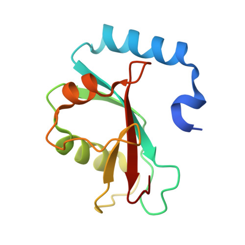
Structural basis of the autophagy-related LC3/Atg13 LIR complex: recognition and interaction mechanism.

Suzuki, H.Tabata, K.Morita, E.Kawasaki, M.Kato, R.Dobson, R.C.Yoshimori, T.Wakatsuki, S.

(2014) Structure 22: 47-58

  • DOI: https://doi.org/10.1016/j.str.2013.09.023
  • Primary Citation of Related Structures:  
    3WAL, 3WAM, 3WAN, 3WAO, 3WAP

  • PubMed Abstract: 

    Autophagy is a bulk degradation pathway that removes cytosolic materials to maintain cellular homeostasis. The autophagy-related gene 13 (Atg13) and microtubule associate protein 1 light chain 3 (LC3) proteins are required for autophagosome formation. We demonstrate that each of the human LC3 isoforms (LC3A, LC3B, and LC3C) interacts with Atg13 via the LC3 interacting region (LIR) of Atg13. Using X-ray crystallography, we solved the macromolecular structures of LC3A and LC3C, along with the complex structures of the LC3 isoforms with the Atg13 LIR. Together, our structural and binding analyses reveal that the side-chain of Lys49 of LC3 acts as a gatekeeper to regulate binding of the LIR. We verified this observation by mutation of Lys49 in LC3A, which significantly reduces LC3A positive puncta formation in cultured cells. Our results suggest that specific affinity of the LC3 isoforms to the Atg13 LIR is required for proper autophagosome formation.


  • Organizational Affiliation

    Biomolecular Interaction Centre, School of Biological Sciences, University of Canterbury, Christchurch 8020, New Zealand; Structural Biology Research Center, Photon Factory, Institute of Materials Structure Science, High Energy Accelerator Research Organization (KEK), Tsukuba, Ibaraki 305-0801, Japan. Electronic address: hironori.suzuki@canterbury.ac.nz.


Macromolecules
Find similar proteins by:  (by identity cutoff)  |  3D Structure
Entity ID: 1
MoleculeChains Sequence LengthOrganismDetailsImage
Microtubule-associated proteins 1A/1B light chain 3C120Homo sapiensMutation(s): 0 
Gene Names: MAP1LC3C
UniProt & NIH Common Fund Data Resources
Find proteins for Q9BXW4 (Homo sapiens)
Explore Q9BXW4 
Go to UniProtKB:  Q9BXW4
PHAROS:  Q9BXW4
GTEx:  ENSG00000197769 
Entity Groups  
Sequence Clusters30% Identity50% Identity70% Identity90% Identity95% Identity100% Identity
UniProt GroupQ9BXW4
Sequence Annotations
Expand
  • Reference Sequence
Small Molecules
Ligands 1 Unique
IDChains Name / Formula / InChI Key2D Diagram3D Interactions
CIT
Query on CIT

Download Ideal Coordinates CCD File 
B [auth A]CITRIC ACID
C6 H8 O7
KRKNYBCHXYNGOX-UHFFFAOYSA-N
Experimental Data & Validation

Experimental Data

  • Method: X-RAY DIFFRACTION
  • Resolution: 1.75 Å
  • R-Value Free: 0.211 
  • R-Value Work: 0.172 
  • R-Value Observed: 0.174 
  • Space Group: P 31 2 1
Unit Cell:
Length ( Å )Angle ( ˚ )
a = 71.72α = 90
b = 71.72β = 90
c = 55.65γ = 120
Software Package:
Software NamePurpose
MOLREPphasing
REFMACrefinement
MOSFLMdata reduction
SCALAdata scaling

Structure Validation

View Full Validation Report


Entry History 

Deposition Data

Revision History  (Full details and data files)

  • Version 1.0: 2013-12-25
    Type: Initial release
  • Version 1.1: 2014-03-12
    Changes: Database references
  • Version 1.2: 2023-11-08
    Changes: Data collection, Database references, Derived calculations, Refinement description