3D5O

Structural recognition and functional activation of FcrR by innate pentraxins


Experimental Data Snapshot

  • Method: X-RAY DIFFRACTION
  • Resolution: 2.80 Å
  • R-Value Free: 0.279 
  • R-Value Work: 0.207 
  • R-Value Observed: 0.221 

wwPDB Validation   3D Report Full Report


Ligand Structure Quality Assessment 


This is version 1.2 of the entry. See complete history


Literature

Structural recognition and functional activation of FcgammaR by innate pentraxins.

Lu, J.Marnell, L.L.Marjon, K.D.Mold, C.Du Clos, T.W.Sun, P.D.

(2008) Nature 456: 989-992

  • DOI: https://doi.org/10.1038/nature07468
  • Primary Citation of Related Structures:  
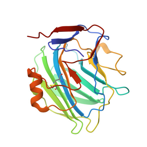
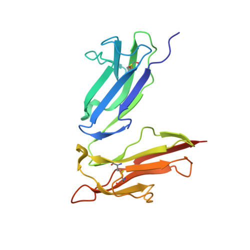
    3D5O

  • PubMed Abstract: 

    Pentraxins are a family of ancient innate immune mediators conserved throughout evolution. The classical pentraxins include serum amyloid P component (SAP) and C-reactive protein, which are two of the acute-phase proteins synthesized in response to infection. Both recognize microbial pathogens and activate the classical complement pathway through C1q (refs 3 and 4). More recently, members of the pentraxin family were found to interact with cell-surface Fcgamma receptors (FcgammaR) and activate leukocyte-mediated phagocytosis. Here we describe the structural mechanism for pentraxin's binding to FcgammaR and its functional activation of FcgammaR-mediated phagocytosis and cytokine secretion. The complex structure between human SAP and FcgammaRIIa reveals a diagonally bound receptor on each SAP pentamer with both D1 and D2 domains of the receptor contacting the ridge helices from two SAP subunits. The 1:1 stoichiometry between SAP and FcgammaRIIa infers the requirement for multivalent pathogen binding for receptor aggregation. Mutational and binding studies show that pentraxins are diverse in their binding specificity for FcgammaR isoforms but conserved in their recognition structure. The shared binding site for SAP and IgG results in competition for FcgammaR binding and the inhibition of immune-complex-mediated phagocytosis by soluble pentraxins. These results establish antibody-like functions for pentraxins in the FcgammaR pathway, suggest an evolutionary overlap between the innate and adaptive immune systems, and have new therapeutic implications for autoimmune diseases.


  • Organizational Affiliation

    Structural Immunology Section, Laboratory of Immunogenetics, National Institute of Allergy and Infectious Diseases, National Institutes of Health, Rockville, Maryland 20852, USA.


Macromolecules
Find similar proteins by:  (by identity cutoff)  |  3D Structure
Entity ID: 1
MoleculeChains Sequence LengthOrganismDetailsImage
Serum amyloid P-component
A, B, C, D, E
204Homo sapiensMutation(s): 0 
UniProt & NIH Common Fund Data Resources
Find proteins for P02743 (Homo sapiens)
Explore P02743 
Go to UniProtKB:  P02743
PHAROS:  P02743
GTEx:  ENSG00000132703 
Entity Groups  
Sequence Clusters30% Identity50% Identity70% Identity90% Identity95% Identity100% Identity
UniProt GroupP02743
Sequence Annotations
Expand
  • Reference Sequence
Find similar proteins by:  (by identity cutoff)  |  3D Structure
Entity ID: 2
MoleculeChains Sequence LengthOrganismDetailsImage
Low affinity immunoglobulin gamma Fc region receptor II-a177Homo sapiensMutation(s): 0 
Gene Names: FCGR2ACD32FCG2FCGR2A1IGFR2
UniProt & NIH Common Fund Data Resources
Find proteins for P12318 (Homo sapiens)
Explore P12318 
Go to UniProtKB:  P12318
PHAROS:  P12318
GTEx:  ENSG00000143226 
Entity Groups  
Sequence Clusters30% Identity50% Identity70% Identity90% Identity95% Identity100% Identity
UniProt GroupP12318
Sequence Annotations
Expand
  • Reference Sequence
Small Molecules
Ligands 3 Unique
IDChains Name / Formula / InChI Key2D Diagram3D Interactions
NAG
Query on NAG

Download Ideal Coordinates CCD File 
J [auth A],
N [auth B],
R [auth C],
V [auth D],
Z [auth E]
2-acetamido-2-deoxy-beta-D-glucopyranose
C8 H15 N O6
OVRNDRQMDRJTHS-FMDGEEDCSA-N
SO4
Query on SO4

Download Ideal Coordinates CCD File 
AA [auth F]
G [auth A]
H [auth A]
I [auth A]
K [auth B]
AA [auth F],
G [auth A],
H [auth A],
I [auth A],
K [auth B],
L [auth B],
M [auth B],
O [auth C],
P [auth C],
Q [auth C],
S [auth D],
T [auth D],
U [auth D],
W [auth E],
X [auth E],
Y [auth E]
SULFATE ION
O4 S
QAOWNCQODCNURD-UHFFFAOYSA-L
GOL
Query on GOL

Download Ideal Coordinates CCD File 
BA [auth F]GLYCEROL
C3 H8 O3
PEDCQBHIVMGVHV-UHFFFAOYSA-N
Experimental Data & Validation

Experimental Data

  • Method: X-RAY DIFFRACTION
  • Resolution: 2.80 Å
  • R-Value Free: 0.279 
  • R-Value Work: 0.207 
  • R-Value Observed: 0.221 
  • Space Group: P 21 21 21
Unit Cell:
Length ( Å )Angle ( ˚ )
a = 64.197α = 90
b = 143.477β = 90
c = 161.588γ = 90
Software Package:
Software NamePurpose
HKL-2000data collection
PHASESphasing
CNSrefinement
HKL-2000data reduction
HKL-2000data scaling

Structure Validation

View Full Validation Report


Ligand Structure Quality Assessment 


Entry History 

Deposition Data

Revision History  (Full details and data files)

  • Version 1.0: 2008-11-11
    Type: Initial release
  • Version 1.1: 2011-07-13
    Changes: Non-polymer description, Version format compliance
  • Version 1.2: 2020-07-29
    Type: Remediation
    Reason: Carbohydrate remediation
    Changes: Data collection, Database references, Derived calculations, Structure summary