2ZY6

Crystal structure of a truncated tRNA, TPHE39A


Experimental Data Snapshot

  • Method: X-RAY DIFFRACTION
  • Resolution: 1.75 Å
  • R-Value Free: 0.240 
  • R-Value Work: 0.207 
  • R-Value Observed: 0.209 

wwPDB Validation   3D Report Full Report


This is version 1.2 of the entry. See complete history


Literature

Deduced RNA binding mechanism of ThiI based on structural and binding analyses of a minimal RNA ligand

Tanaka, Y.Yamagata, S.Kitago, Y.Yamada, Y.Chimnaronk, S.Yao, M.Tanaka, I.

(2009) RNA 15: 1498-1506

  • DOI: https://doi.org/10.1261/rna.1614709
  • Primary Citation of Related Structures:  
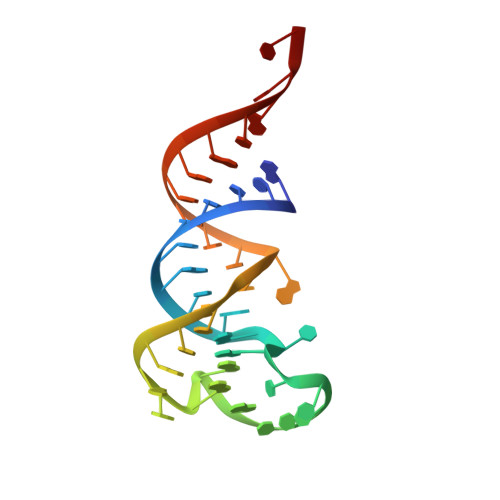
    2ZY6

  • PubMed Abstract: 

    ThiI catalyzes the thio-introduction reaction to tRNA, and a truncated tRNA consisting of 39 nucleotides, TPHE39A, is the minimal RNA substrate for modification by ThiI from Escherichia coli. To examine the molecular basis of the tRNA recognition by ThiI, we have solved the crystal structure of TPHE39A, which showed that base pairs in the T-stem were almost completely disrupted, although those in the acceptor-stem were preserved. Gel shift assays and isothermal titration calorimetry experiments showed that ThiI can efficiently bind with not only tRNA(Phe) but also TPHE39A. Binding assays using truncated ThiI, i.e., N- and C-terminal domains of ThiI, showed that the N-domain can bind with both tRNA(Phe) and TPHE39A, whereas the C-domain cannot. These results indicated that the N-domain of ThiI recognizes the acceptor-stem region. Thermodynamic analysis indicated that the C-domain also affects RNA binding by its enthalpically favorable, but entropically unfavorable, contribution. In addition, circular dichroism spectra showed that the C-domain induced a conformation change in tRNA(Phe). Based on these results, a possible RNA binding mechanism of ThiI in which the N-terminal domain recognizes the acceptor-stem region and the C-terminal region causes a conformational change of RNA is proposed.


  • Organizational Affiliation

    Creative Research Institute Sousei, Hokkaido University, Sapporo, 001-0021, Japan. tanaka@castor.sci.hokudai.ac.jp


Macromolecules
Find similar nucleic acids by:  (by identity cutoff)  |  3D Structure
Entity ID: 1
MoleculeChains LengthOrganismImage
phenylalanyl transfer RNA39N/A
Sequence Annotations
Expand
  • Reference Sequence
Small Molecules
Ligands 3 Unique
IDChains Name / Formula / InChI Key2D Diagram3D Interactions
CA
Query on CA

Download Ideal Coordinates CCD File 
D [auth A]
E [auth A]
F [auth A]
G [auth A]
H [auth A]
D [auth A],
E [auth A],
F [auth A],
G [auth A],
H [auth A],
I [auth A],
J [auth A]
CALCIUM ION
Ca
BHPQYMZQTOCNFJ-UHFFFAOYSA-N
CL
Query on CL

Download Ideal Coordinates CCD File 
C [auth A]CHLORIDE ION
Cl
VEXZGXHMUGYJMC-UHFFFAOYSA-M
MG
Query on MG

Download Ideal Coordinates CCD File 
B [auth A]MAGNESIUM ION
Mg
JLVVSXFLKOJNIY-UHFFFAOYSA-N
Experimental Data & Validation

Experimental Data

  • Method: X-RAY DIFFRACTION
  • Resolution: 1.75 Å
  • R-Value Free: 0.240 
  • R-Value Work: 0.207 
  • R-Value Observed: 0.209 
  • Space Group: P 61 2 2
Unit Cell:
Length ( Å )Angle ( ˚ )
a = 62.366α = 90
b = 62.366β = 90
c = 115.2γ = 120
Software Package:
Software NamePurpose
REFMACrefinement
HKL-2000data collection
HKL-2000data reduction
HKL-2000data scaling
SHELXEmodel building

Structure Validation

View Full Validation Report


Entry History 

Deposition Data

Revision History  (Full details and data files)

  • Version 1.0: 2009-06-30
    Type: Initial release
  • Version 1.1: 2011-07-13
    Changes: Version format compliance
  • Version 1.2: 2024-03-13
    Changes: Data collection, Database references, Derived calculations