2X98

H.SALINARUM ALKALINE PHOSPHATASE


Experimental Data Snapshot

  • Method: X-RAY DIFFRACTION
  • Resolution: 1.70 Å
  • R-Value Free: 0.198 
  • R-Value Work: 0.160 
  • R-Value Observed: 0.163 

wwPDB Validation   3D Report Full Report


This is version 1.1 of the entry. See complete history


Literature

Structural and Biochemical Characterization of a Halophilic Archaeal Alkaline Phosphatase.

Wende, A.Johansson, P.Vollrath, R.Dyall-Smith, M.Oesterhelt, D.Grininger, M.

(2010) J Mol Biol 400: 52

  • DOI: https://doi.org/10.1016/j.jmb.2010.04.057
  • Primary Citation of Related Structures:  
    2X98

  • PubMed Abstract: 

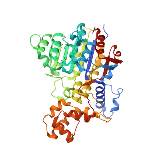
    Phosphate is an essential component of all cells that must be taken up from the environment. Prokaryotes commonly secrete alkaline phosphatases (APs) to recruit phosphate from organic compounds by hydrolysis. In this study, the AP from Halobacterium salinarum, an archaeon that lives in a saturated salt environment, has been functionally and structurally characterized. The core fold and the active-site architecture of the H. salinarum enzyme are similar to other AP structures. These generally form dimers composed of dominant beta-sheet structures sandwiched by alpha-helices and have well-accessible active sites. The surface of the enzyme is predicted to be highly negatively charged, like other proteins of extreme halophiles. In addition to the conserved core, most APs contain a crown domain that strongly varies within species. In the H. salinarum AP, the crown domain is made of an acyl-carrier-protein-like fold. Different from other APs, it is not involved in dimer formation. We compare the archaeal AP with its bacterial and eukaryotic counterparts, and we focus on the role of crown domains in enhancing protein stability, regulating enzyme function, and guiding phosphoesters into the active-site funnel.


  • Organizational Affiliation

    Department of Membrane Biochemistry, Max Planck Institute of Biochemistry, Am Klopferspitz 18, 82152 Martinsried, Germany.


Macromolecules
Find similar proteins by:  (by identity cutoff)  |  3D Structure
Entity ID: 1
MoleculeChains Sequence LengthOrganismDetailsImage
ALKALINE PHOSPHATASE
A, B
431Halobacterium salinarum R1Mutation(s): 0 
EC: 3.1.3.1
UniProt
Find proteins for B0R9W3 (Halobacterium salinarum (strain ATCC 29341 / DSM 671 / R1))
Explore B0R9W3 
Go to UniProtKB:  B0R9W3
Entity Groups  
Sequence Clusters30% Identity50% Identity70% Identity90% Identity95% Identity100% Identity
UniProt GroupB0R9W3
Sequence Annotations
Expand
  • Reference Sequence
Small Molecules
Ligands 5 Unique
IDChains Name / Formula / InChI Key2D Diagram3D Interactions
PO4
Query on PO4

Download Ideal Coordinates CCD File 
C [auth A],
R [auth B]
PHOSPHATE ION
O4 P
NBIIXXVUZAFLBC-UHFFFAOYSA-K
ZN
Query on ZN

Download Ideal Coordinates CCD File 
D [auth A],
E [auth A],
S [auth B],
T [auth B]
ZINC ION
Zn
PTFCDOFLOPIGGS-UHFFFAOYSA-N
CL
Query on CL

Download Ideal Coordinates CCD File 
BA [auth B]
CA [auth B]
DA [auth B]
EA [auth B]
FA [auth B]
BA [auth B],
CA [auth B],
DA [auth B],
EA [auth B],
FA [auth B],
GA [auth B],
HA [auth B],
L [auth A],
M [auth A],
N [auth A],
O [auth A],
P [auth A],
Q [auth A]
CHLORIDE ION
Cl
VEXZGXHMUGYJMC-UHFFFAOYSA-M
MG
Query on MG

Download Ideal Coordinates CCD File 
F [auth A]
G [auth A]
H [auth A]
I [auth A]
J [auth A]
F [auth A],
G [auth A],
H [auth A],
I [auth A],
J [auth A],
U [auth B],
V [auth B],
W [auth B],
X [auth B],
Y [auth B],
Z [auth B]
MAGNESIUM ION
Mg
JLVVSXFLKOJNIY-UHFFFAOYSA-N
NA
Query on NA

Download Ideal Coordinates CCD File 
AA [auth B],
K [auth A]
SODIUM ION
Na
FKNQFGJONOIPTF-UHFFFAOYSA-N
Experimental Data & Validation

Experimental Data

  • Method: X-RAY DIFFRACTION
  • Resolution: 1.70 Å
  • R-Value Free: 0.198 
  • R-Value Work: 0.160 
  • R-Value Observed: 0.163 
  • Space Group: C 1 2 1
Unit Cell:
Length ( Å )Angle ( ˚ )
a = 72.143α = 90
b = 152.524β = 110
c = 86.799γ = 90
Software Package:
Software NamePurpose
REFMACrefinement
MOSFLMdata reduction
SCALEPACKdata scaling
PHASERphasing

Structure Validation

View Full Validation Report


Entry History 

Deposition Data

Revision History  (Full details and data files)

  • Version 1.0: 2010-05-19
    Type: Initial release
  • Version 1.1: 2012-10-03
    Changes: Derived calculations, Refinement description, Version format compliance