2WG3

Crystal structure of the complex between human hedgehog-interacting protein HIP and desert hedgehog without calcium


Experimental Data Snapshot

  • Method: X-RAY DIFFRACTION
  • Resolution: 2.60 Å
  • R-Value Free: 0.281 
  • R-Value Work: 0.224 
  • R-Value Observed: 0.227 

wwPDB Validation   3D Report Full Report


Ligand Structure Quality Assessment 


This is version 1.3 of the entry. See complete history


Literature

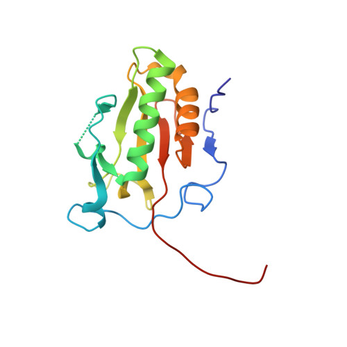
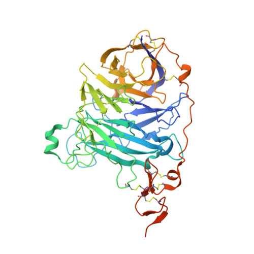
Structural Insights Into Hedgehog Ligand Sequestration by the Human Hedgehog-Interacting Protein Hip

Bishop, B.Aricescu, A.R.Harlos, K.O'Callaghan, C.A.Jones, E.Y.Siebold, C.

(2009) Nat Struct Mol Biol 16: 698

  • DOI: https://doi.org/10.1038/nsmb.1607
  • Primary Citation of Related Structures:  
    2WFQ, 2WFR, 2WFT, 2WFX, 2WG3, 2WG4

  • PubMed Abstract: 

    Hedgehog (Hh) morphogens have fundamental roles in development, whereas dysregulation of Hh signaling leads to disease. Multiple cell-surface receptors are responsible for transducing and/or regulating Hh signals. Among these, the Hedgehog-interacting protein (Hhip) is a highly conserved, vertebrate-specific inhibitor of Hh signaling. We have solved a series of crystal structures for the human HHIP ectodomain and Desert hedgehog (DHH) in isolation, as well as HHIP in complex with DHH (HHIP-DHH) and Sonic hedgehog (Shh) (HHIP-Shh), with and without Ca2+. The interaction determinants, confirmed by biophysical studies and mutagenesis, reveal previously uncharacterized and distinct functions for the Hh Zn2+ and Ca2+ binding sites--functions that may be common to all vertebrate Hh proteins. Zn2+ makes a key contribution to the Hh-HHIP interface, whereas Ca2+ is likely to prevent electrostatic repulsion between the two proteins, suggesting an important modulatory role. This interplay of several metal binding sites suggests a tuneable mechanism for regulation of Hh signaling.


  • Organizational Affiliation

    Division of Structural Biology, Wellcome Trust Centre for Human Genetics, University of Oxford, Oxford, UK.


Macromolecules
Find similar proteins by:  (by identity cutoff)  |  3D Structure
Entity ID: 1
MoleculeChains Sequence LengthOrganismDetailsImage
DESERT HEDGEHOG PROTEIN N-PRODUCT
A, B
164Homo sapiensMutation(s): 0 
UniProt & NIH Common Fund Data Resources
Find proteins for O43323 (Homo sapiens)
Explore O43323 
Go to UniProtKB:  O43323
PHAROS:  O43323
GTEx:  ENSG00000139549 
Entity Groups  
Sequence Clusters30% Identity50% Identity70% Identity90% Identity95% Identity100% Identity
UniProt GroupO43323
Sequence Annotations
Expand
  • Reference Sequence
Find similar proteins by:  (by identity cutoff)  |  3D Structure
Entity ID: 2
MoleculeChains Sequence LengthOrganismDetailsImage
HEDGEHOG-INTERACTING PROTEIN
C, D
463Homo sapiensMutation(s): 0 
UniProt & NIH Common Fund Data Resources
Find proteins for Q96QV1 (Homo sapiens)
Explore Q96QV1 
Go to UniProtKB:  Q96QV1
PHAROS:  Q96QV1
GTEx:  ENSG00000164161 
Entity Groups  
Sequence Clusters30% Identity50% Identity70% Identity90% Identity95% Identity100% Identity
UniProt GroupQ96QV1
Sequence Annotations
Expand
  • Reference Sequence
Small Molecules
Ligands 4 Unique
IDChains Name / Formula / InChI Key2D Diagram3D Interactions
NAG
Query on NAG

Download Ideal Coordinates CCD File 
I [auth C],
M [auth D]
2-acetamido-2-deoxy-beta-D-glucopyranose
C8 H15 N O6
OVRNDRQMDRJTHS-FMDGEEDCSA-N
PO4
Query on PO4

Download Ideal Coordinates CCD File 
H [auth C],
L [auth D]
PHOSPHATE ION
O4 P
NBIIXXVUZAFLBC-UHFFFAOYSA-K
ZN
Query on ZN

Download Ideal Coordinates CCD File 
E [auth A],
G [auth B]
ZINC ION
Zn
PTFCDOFLOPIGGS-UHFFFAOYSA-N
CL
Query on CL

Download Ideal Coordinates CCD File 
F [auth A]
J [auth C]
K [auth C]
N [auth D]
O [auth D]
F [auth A],
J [auth C],
K [auth C],
N [auth D],
O [auth D],
P [auth D]
CHLORIDE ION
Cl
VEXZGXHMUGYJMC-UHFFFAOYSA-M
Experimental Data & Validation

Experimental Data

  • Method: X-RAY DIFFRACTION
  • Resolution: 2.60 Å
  • R-Value Free: 0.281 
  • R-Value Work: 0.224 
  • R-Value Observed: 0.227 
  • Space Group: P 21 21 21
Unit Cell:
Length ( Å )Angle ( ˚ )
a = 102.821α = 90
b = 110.701β = 90
c = 144.381γ = 90
Software Package:
Software NamePurpose
REFMACrefinement
HKL-2000data reduction
SCALEPACKdata scaling
PHASERphasing

Structure Validation

View Full Validation Report


Ligand Structure Quality Assessment 


Entry History 

Deposition Data

Revision History  (Full details and data files)

  • Version 1.0: 2009-06-30
    Type: Initial release
  • Version 1.1: 2011-07-13
    Changes: Advisory, Refinement description, Version format compliance
  • Version 1.2: 2020-07-29
    Type: Remediation
    Reason: Carbohydrate remediation
    Changes: Data collection, Derived calculations, Other, Structure summary
  • Version 1.3: 2023-12-13
    Changes: Data collection, Database references, Refinement description, Structure summary