2O6N

RH4B: designed right-handed coiled coil tetramer with all biological amino acids


Experimental Data Snapshot

  • Method: X-RAY DIFFRACTION
  • Resolution: 1.10 Å
  • R-Value Free: 0.132 
  • R-Value Work: 0.115 
  • R-Value Observed: 0.116 

wwPDB Validation   3D Report Full Report


This is version 1.2 of the entry. See complete history


Literature

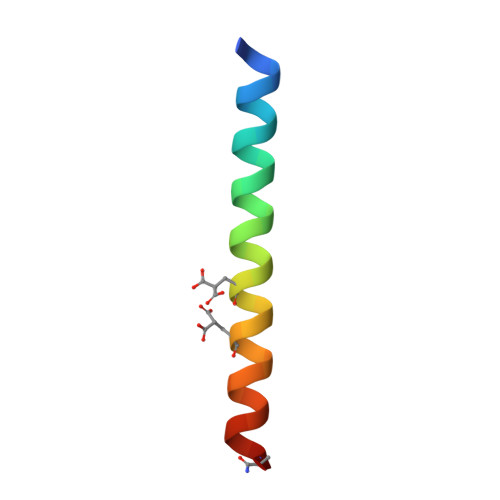
Structure of a designed, right-handed coiled-coil tetramer containing all biological amino acids.

Sales, M.Plecs, J.J.Holton, J.M.Alber, T.

(2007) Protein Sci 16: 2224-2232

  • DOI: https://doi.org/10.1110/ps.062702907
  • Primary Citation of Related Structures:  
    2O6N

  • PubMed Abstract: 

    The previous design of an unprecedented family of two-, three-, and four-helical, right-handed coiled coils utilized nonbiological amino acids to efficiently pack spaces in the oligomer cores. Here we show that a stable, right-handed parallel tetrameric coiled coil, called RH4B, can be designed entirely using biological amino acids. The X-ray crystal structure of RH4B was determined to 1.1 Angstrom resolution using a designed metal binding site to coordinate a single Yb(2+) ion per 33-amino acid polypeptide chain. The resulting experimental phases were particularly accurate, and the experimental electron density map provided an especially clear, unbiased view of the molecule. The RH4B structure closely matched the design, with equivalent core rotamers and an overall root-mean-square deviation for the N-terminal repeat of the tetramer of 0.24 Angstrom. The clarity and resolution of the electron density map, however, revealed alternate rotamers and structural differences between the three sequence repeats in the molecule. These results suggest that the RH4B structure populates an unanticipated variety of structures.


  • Organizational Affiliation

    Department of Physics, University of California, Berkeley, California 94720-3206, USA.


Macromolecules
Find similar proteins by:  (by identity cutoff)  |  3D Structure
Entity ID: 1
MoleculeChains Sequence LengthOrganismDetailsImage
RH4B designed peptide35N/AMutation(s): 0 
Entity Groups  
Sequence Clusters30% Identity50% Identity70% Identity90% Identity95% Identity100% Identity
Sequence Annotations
Expand
  • Reference Sequence
Small Molecules
Ligands 1 Unique
IDChains Name / Formula / InChI Key2D Diagram3D Interactions
YB2
Query on YB2

Download Ideal Coordinates CCD File 
B [auth A]YTTERBIUM (II) ION
Yb
SVJLBBSKJLMTRQ-UHFFFAOYSA-N
Modified Residues  1 Unique
IDChains TypeFormula2D DiagramParent
CGU
Query on CGU
A
L-PEPTIDE LINKINGC6 H9 N O6GLU
Experimental Data & Validation

Experimental Data

  • Method: X-RAY DIFFRACTION
  • Resolution: 1.10 Å
  • R-Value Free: 0.132 
  • R-Value Work: 0.115 
  • R-Value Observed: 0.116 
  • Space Group: P 4 21 2
Unit Cell:
Length ( Å )Angle ( ˚ )
a = 38.356α = 90
b = 38.356β = 90
c = 49.679γ = 90
Software Package:
Software NamePurpose
REFMACrefinement
Blu-Icedata collection
MOSFLMdata reduction
SCALAdata scaling
SOLVEphasing

Structure Validation

View Full Validation Report


Entry History 

Deposition Data

Revision History  (Full details and data files)

  • Version 1.0: 2007-10-23
    Type: Initial release
  • Version 1.1: 2011-07-13
    Changes: Derived calculations, Version format compliance
  • Version 1.2: 2023-12-27
    Changes: Data collection, Database references, Derived calculations