2NTZ

Structure of a ParB-DNA complex reveals a double B-box interaction


Experimental Data Snapshot

  • Method: X-RAY DIFFRACTION
  • Resolution: 3.35 Å
  • R-Value Free: 0.336 
  • R-Value Work: 0.288 
  • R-Value Observed: 0.288 

wwPDB Validation   3D Report Full Report


This is version 1.4 of the entry. See complete history


Literature

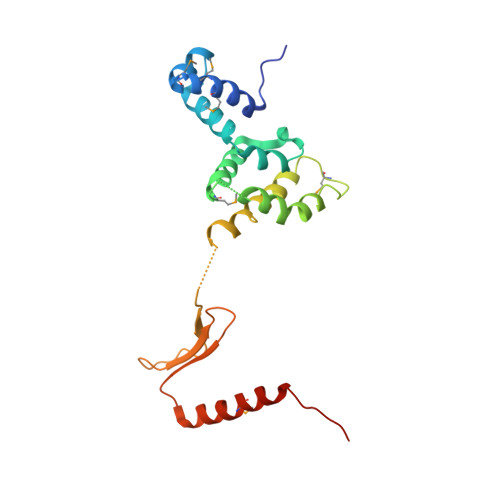
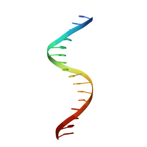
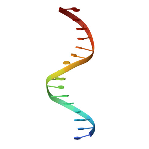
Structure of a four-way bridged ParB-DNA complex provides insight into P1 segrosome assembly.

Schumacher, M.A.Mansoor, A.Funnell, B.E.

(2007) J Biol Chem 282: 10456-10464

  • DOI: https://doi.org/10.1074/jbc.M610603200
  • Primary Citation of Related Structures:  
    2NTZ

  • PubMed Abstract: 

    The plasmid partition process is essential for plasmid propagation and is mediated by par systems, consisting of centromere-like sites and two proteins, ParA and ParB. In the first step of partition by the archetypical P1 system, ParB binds a complicated centromere-like site to form a large nucleoprotein segrosome. ParB is a dimeric DNA-binding protein that can bridge between both A-boxes and B-boxes located on the centromere. Its helix-turn-helix domains bind A-boxes and the dimer domain binds B-boxes. Binding of the first ParB dimer nucleates the remaining ParB molecules onto the centromere site, which somehow leads to the formation of a condensed segrosome superstructure. To further understand this unique DNA spreading capability of ParB, we crystallized and determined the structure of a 1:2 ParB-(142-333):A3-B2-box complex to 3.35A resolution. The structure reveals a remarkable four-way, protein-DNA bridged complex in which both ParB helix-turn-helix domains simultaneously bind adjacent A-boxes and the dimer domain bridges between two B-boxes. The multibridging capability and the novel dimer domain-B-box interaction, which juxtaposes the DNA sites close in space, suggests a mechanism for the formation of the wrapped solenoid-like segrosome superstructure. This multibridging capability of ParB is likely critical in its partition complex formation and pairing functions.


  • Organizational Affiliation

    Department of Biochemistry and Molecular Biology, University of Texas M. D. Anderson Cancer Center, Houston, Texas 77030, USA. maschuma@mdanderson


Macromolecules

Find similar proteins by:  (by identity cutoff)  |  3D Structure
Entity ID: 3
MoleculeChains Sequence LengthOrganismDetailsImage
ParBE [auth A],
F [auth B]
192Punavirus P1Mutation(s): 6 
Gene Names: parb
UniProt
Find proteins for Q38420 (Punavirus P1)
Explore Q38420 
Go to UniProtKB:  Q38420
Entity Groups  
Sequence Clusters30% Identity50% Identity70% Identity90% Identity95% Identity100% Identity
UniProt GroupQ38420
Sequence Annotations
Expand
  • Reference Sequence

Find similar nucleic acids by:  Sequence   |   3D Structure  

Entity ID: 1
MoleculeChains LengthOrganismImage
5'-D(*CP*GP*TP*GP*AP*AP*AP*TP*CP*GP*CP*CP*AP*CP*GP*A)-3'A [auth Y],
C [auth E]
16N/A
Sequence Annotations
Expand
  • Reference Sequence

Find similar nucleic acids by:  Sequence   |   3D Structure  

Entity ID: 2
MoleculeChains LengthOrganismImage
5'-D(*TP*CP*GP*TP*GP*GP*CP*GP*AP*TP*TP*TP*CP*AP*CP*G)-3'B [auth W],
D [auth U]
16N/A
Sequence Annotations
Expand
  • Reference Sequence
Small Molecules
Modified Residues  1 Unique
IDChains TypeFormula2D DiagramParent
MSE
Query on MSE
E [auth A],
F [auth B]
L-PEPTIDE LINKINGC5 H11 N O2 SeMET
Experimental Data & Validation

Experimental Data

  • Method: X-RAY DIFFRACTION
  • Resolution: 3.35 Å
  • R-Value Free: 0.336 
  • R-Value Work: 0.288 
  • R-Value Observed: 0.288 
  • Space Group: P 31 2 1
Unit Cell:
Length ( Å )Angle ( ˚ )
a = 144.42α = 90
b = 144.42β = 90
c = 78.88γ = 120
Software Package:
Software NamePurpose
CNSrefinement
ADSCdata collection
MOSFLMdata reduction
CCP4data scaling
EPMRphasing

Structure Validation

View Full Validation Report


Entry History 

Deposition Data

Revision History  (Full details and data files)

  • Version 1.0: 2007-02-20
    Type: Initial release
  • Version 1.1: 2008-05-01
    Changes: Version format compliance
  • Version 1.2: 2011-07-13
    Changes: Version format compliance
  • Version 1.3: 2023-08-30
    Changes: Data collection, Database references, Derived calculations, Refinement description
  • Version 1.4: 2023-11-15
    Changes: Data collection