2JXR

STRUCTURE OF YEAST PROTEINASE A


Experimental Data Snapshot

  • Method: X-RAY DIFFRACTION
  • Resolution: 2.40 Å
  • R-Value Free: 0.270 
  • R-Value Work: 0.193 

wwPDB Validation   3D Report Full Report


This is version 2.1 of the entry. See complete history


Literature

The three-dimensional structure at 2.4 A resolution of glycosylated proteinase A from the lysosome-like vacuole of Saccharomyces cerevisiae.

Aguilar, C.F.Cronin, N.B.Badasso, M.Dreyer, T.Newman, M.P.Cooper, J.B.Hoover, D.J.Wood, S.P.Johnson, M.S.Blundell, T.L.

(1997) J Mol Biol 267: 899-915

  • DOI: https://doi.org/10.1006/jmbi.1996.0880
  • Primary Citation of Related Structures:  
    2JXR

  • PubMed Abstract: 

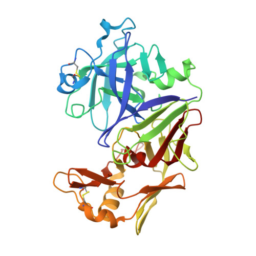
    The crystal structures of glycosylated native proteinase A, an aspartic proteinase found in the vacuole of Saccharomyces cerevisiae, and its complex with a difluorostatone-containing tripeptide have been determined by molecular replacement to 3.5 A and 2.4 A resolutions, respectively. Superposition of the bound and native forms gave an r.m.s. difference of 0.6 A largely reflecting the poor resolution of the native crystal structure. The secondary and tertiary structures are highly similar to those found in porcine pepsin and lysosomal cathepsin D; superposition of the structure of proteinase A bound to the difluorostatone inhibitor on those of pepsin and cathepsin D gave pairwise r.m.s. differences for C(alpha) atoms of 1.36 A and 0.88 A. Most differences occur in loop regions. Comparison of the structure of the proteinase A-difluorostatone complex with that of endothiapepsin bound with the same inhibitor shows that the conformation and hydrogen bond interactions of the inhibitor in the active site are very similar, even though the enzymes have only 27% sequence identity. Electron density for the crystal structure of the proteinase A complex reveals five residues of the oligosaccharide structure attached to Asn67: Man-(1 --> 2)-alpha-Man-(1 --> 3)-beta-Man-(1 --> 4)-beta-GlcNAc-(1 --> 4)-beta-GlcNAc-Asn-67. The first three residues of the oligosaccharide cover the same region of the protein surface as those of the oligosaccharide attached to the equivalent position in cathepsin D. The second carbohydrate attachment site is disordered beyond the first carbohydrate residue in both enzymes.


  • Organizational Affiliation

    Department of Crystallography, Birkbeck College, London, UK.


Macromolecules
Find similar proteins by:  (by identity cutoff)  |  3D Structure
Entity ID: 1
MoleculeChains Sequence LengthOrganismDetailsImage
PROTEINASE A329Saccharomyces cerevisiaeMutation(s): 1 
EC: 3.4.23.25
UniProt
Find proteins for P07267 (Saccharomyces cerevisiae (strain ATCC 204508 / S288c))
Explore P07267 
Go to UniProtKB:  P07267
Entity Groups  
Sequence Clusters30% Identity50% Identity70% Identity90% Identity95% Identity100% Identity
UniProt GroupP07267
Sequence Annotations
Expand
  • Reference Sequence
Oligosaccharides

Help

Entity ID: 2
MoleculeChains Length2D Diagram Glycosylation3D Interactions
alpha-D-mannopyranose-(1-2)-beta-D-mannopyranose-(1-3)-beta-D-mannopyranose-(1-4)-2-acetamido-2-deoxy-beta-D-glucopyranose-(1-4)-2-acetamido-2-deoxy-beta-D-glucopyranose
B
5N-Glycosylation
Glycosylation Resources
GlyTouCan:  G86273CV
GlyCosmos:  G86273CV
GlyGen:  G86273CV
Small Molecules
Ligands 2 Unique
IDChains Name / Formula / InChI Key2D Diagram3D Interactions
2Z3
Query on 2Z3

Download Ideal Coordinates CCD File 
C [auth A]N-(morpholin-4-ylcarbonyl)-L-phenylalanyl-N-[(1R)-1-(cyclohexylmethyl)-3,3-difluoro-2,2-dihydroxy-4-(methylamino)-4-oxobutyl]-L-norleucinamide
C32 H49 F2 N5 O7
IPZOKQNUWWOCTK-GSDHBNRESA-N
NAG
Query on NAG

Download Ideal Coordinates CCD File 
D [auth A]2-acetamido-2-deoxy-beta-D-glucopyranose
C8 H15 N O6
OVRNDRQMDRJTHS-FMDGEEDCSA-N
Binding Affinity Annotations 
IDSourceBinding Affinity
2Z3 Binding MOAD:  2JXR Ki: 90 (nM) from 1 assay(s)
Biologically Interesting Molecules (External Reference) 1 Unique
Entity ID: 3
IDChains NameType/Class2D Diagram3D Interactions
PRD_000416 (2Z3)
Query on PRD_000416
C [auth A]CP-81,282, MOR-PHE-NLE-CHF-NMEPeptide-like / Inhibitor
Experimental Data & Validation

Experimental Data

  • Method: X-RAY DIFFRACTION
  • Resolution: 2.40 Å
  • R-Value Free: 0.270 
  • R-Value Work: 0.193 
  • Space Group: P 32 2 1
Unit Cell:
Length ( Å )Angle ( ˚ )
a = 86.7α = 90
b = 86.7β = 90
c = 110.4γ = 120
Software Package:
Software NamePurpose
WEISdata collection
Agrovatadata reduction
RESTRAINrefinement
WEISdata reduction
Agrovatadata scaling

Structure Validation

View Full Validation Report


Entry History 

Deposition Data

Revision History  (Full details and data files)

  • Version 1.0: 1997-10-29
    Type: Initial release
  • Version 1.1: 2008-03-24
    Changes: Version format compliance
  • Version 1.2: 2011-07-13
    Changes: Atomic model, Database references, Derived calculations, Non-polymer description, Structure summary, Version format compliance
  • Version 1.3: 2012-12-12
    Changes: Other
  • Version 2.0: 2020-07-29
    Type: Remediation
    Reason: Carbohydrate remediation
    Changes: Advisory, Atomic model, Data collection, Derived calculations, Non-polymer description, Other, Refinement description, Structure summary
  • Version 2.1: 2021-11-03
    Changes: Database references, Structure summary