2JAU

Crystal structure of D41N variant of human mitochondrial 5'(3')- deoxyribonucleotidase (mdN) in complex with 3'-azidothymidine 5'- monophosphate


Experimental Data Snapshot

  • Method: X-RAY DIFFRACTION
  • Resolution: 1.80 Å
  • R-Value Free: 0.214 
  • R-Value Work: 0.182 
  • R-Value Observed: 0.183 

wwPDB Validation   3D Report Full Report


Ligand Structure Quality Assessment 


This is version 1.5 of the entry. See complete history


Literature

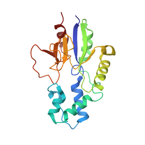
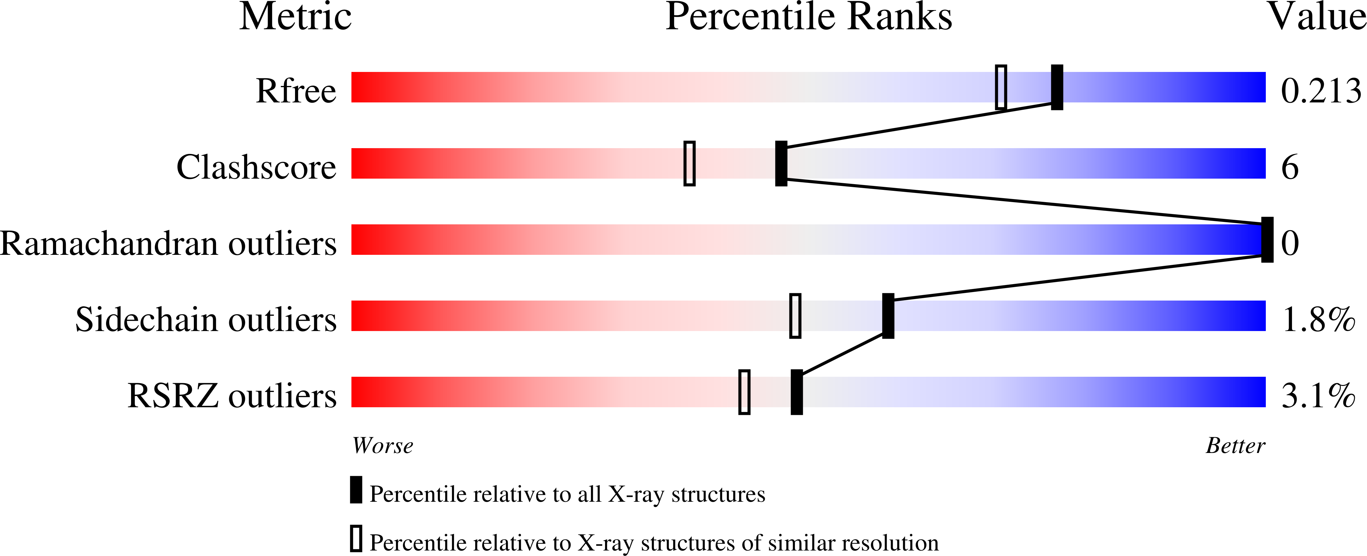
Crystal Structures of Human and Murine Deoxyribonucleotidases: Insights Into Recognition of Substrates and Nucleotide Analogues.

Wallden, K.Rinaldo-Matthis, A.Ruzzenente, B.Rampazzo, C.Bianchi, V.Nordlund, P.

(2007) Biochemistry 46: 13809

  • DOI: https://doi.org/10.1021/bi7014794
  • Primary Citation of Related Structures:  
    2JAO, 2JAR, 2JAU, 2JAW

  • PubMed Abstract: 

    Cytosolic 5'(3')-deoxyribonucleotidase (cdN) and mitochondrial 5'(3')-deoxyribonucleotidase (mdN) catalyze the dephosphorylation of deoxyribonucleoside monophosphates and regulate dTTP formation in cytosol and mitochondria, protecting DNA replication from imbalanced precursor pools. They can also interfere with the phosphorylation-dependent activation of nucleoside analogues used in anticancer and antiviral treatment. To understand the relatively narrow substrate specificity of these two enzymes and their ability to use nucleotide analogues as substrates, we determined the crystal structures of human cdN in complex with deoxyuridine, murine cdN in complex with dUMP and dGMP, and human mdN in complex with the nucleotide analogues AZTMP and BVdUMP. Our results show that the active site residues Leu45 and Tyr65 in cdN form a more favorable binding surface for purine nucleotides than the corresponding Trp75 and Trp76 in mdN, explaining why cdN has higher activity for purine nucleotides than does mdN. The molecular interactions of mdN with AZTMP and BVdUMP indicate why these nucleotide analogues are poorer substrates as compared with the physiological substrate, and they provide a structural rationale for the design of drugs that are less prone to inactivation by the deoxyribonucleotidases. We suggest that introduction of substituents in the 3'-position may result in nucleoside analogues with increased resistance to dephosphorylation.


  • Organizational Affiliation

    Department of Biochemistry and Biophysics, Stockholm University, SE-106 91 Stockholm, Sweden.


Macromolecules
Find similar proteins by:  (by identity cutoff)  |  3D Structure
Entity ID: 1
MoleculeChains Sequence LengthOrganismDetailsImage
5'(3')-DEOXYRIBONUCLEOTIDASE197Homo sapiensMutation(s): 1 
EC: 3.1.3
UniProt & NIH Common Fund Data Resources
Find proteins for Q9NPB1 (Homo sapiens)
Explore Q9NPB1 
Go to UniProtKB:  Q9NPB1
PHAROS:  Q9NPB1
GTEx:  ENSG00000205309 
Entity Groups  
Sequence Clusters30% Identity50% Identity70% Identity90% Identity95% Identity100% Identity
UniProt GroupQ9NPB1
Sequence Annotations
Expand
  • Reference Sequence
Experimental Data & Validation

Experimental Data

  • Method: X-RAY DIFFRACTION
  • Resolution: 1.80 Å
  • R-Value Free: 0.214 
  • R-Value Work: 0.182 
  • R-Value Observed: 0.183 
  • Space Group: P 43 21 2
Unit Cell:
Length ( Å )Angle ( ˚ )
a = 73.59α = 90
b = 73.59β = 90
c = 105.74γ = 90
Software Package:
Software NamePurpose
REFMACrefinement
XDSdata reduction
XSCALEdata scaling
MOLREPphasing

Structure Validation

View Full Validation Report


Ligand Structure Quality Assessment 


Entry History 

Deposition Data

Revision History  (Full details and data files)

  • Version 1.0: 2007-12-11
    Type: Initial release
  • Version 1.1: 2011-05-08
    Changes: Version format compliance
  • Version 1.2: 2011-07-13
    Changes: Version format compliance
  • Version 1.3: 2018-01-17
    Changes: Data collection
  • Version 1.4: 2019-01-30
    Changes: Data collection, Experimental preparation
  • Version 1.5: 2019-02-06
    Changes: Data collection, Experimental preparation