Structural Studies of Thymidine Kinases from Bacillus Anthracis and Bacillus Cereus Provide Insights Into Quaternary Structure and Conformational Changes Upon Substrate Binding
Kosinska, U., Carnrot, C., Sandrini, M.P.B., Clausen, A.R., Wang, L., Piskur, J., Eriksson, S., Eklund, H.(2007) FEBS J 274: 727
- PubMed: 17288553
- DOI: https://doi.org/10.1111/j.1742-4658.2006.05617.x
- Primary Citation of Related Structures:
2J9R, 2JA1 - PubMed Abstract:
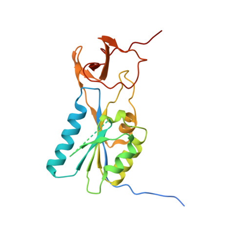
Thymidine kinase (TK) is the key enzyme in salvaging thymidine to produce thymidine monophosphate. Owing to its ability to phosphorylate nucleoside analogue prodrugs, TK has gained attention as a rate-limiting drug activator. We describe the structures of two bacterial TKs, one from the pathogen Bacillus anthracis in complex with the substrate dT, and the second from the food-poison-associated Bacillus cereus in complex with the feedback inhibitor dTTP. Interestingly, in contrast with previous structures of TK in complex with dTTP, in this study dTTP occupies the phosphate donor site and not the phosphate acceptor site. This results in several conformational changes compared with TK structures described previously. One of the differences is the way tetramers are formed. Unlike B. anthracis TK, B. cereus TK shows a loose tetramer. Moreover, the lasso-domain is in open conformation in B. cereus TK without any substrate in the active site, whereas in B. anthracis TK the loop conformation is closed and thymidine occupies the active site. Another conformational difference lies within a region of 20 residues that we refer to as phosphate-binding beta-hairpin. The phosphate-binding beta-hairpin seems to be a flexible region of the enzyme which becomes ordered upon formation of hydrogen bonds to the alpha-phosphate of the phosphate donor, dTTP. In addition to descriptions of the different conformations that TK may adopt during the course of reaction, the oligomeric state of the enzyme is investigated.
Organizational Affiliation:
Department of Molecular Biology, Swedish University of Agricultural Sciences, Uppsala Biomedical Centre, Sweden.