2FBE

Crystal Structure of the PRYSPRY-domain


Experimental Data Snapshot

  • Method: X-RAY DIFFRACTION
  • Resolution: 2.52 Å
  • R-Value Free: 0.277 
  • R-Value Work: 0.227 
  • R-Value Observed: 0.227 

wwPDB Validation   3D Report Full Report


This is version 1.2 of the entry. See complete history


Literature

Structure of the PRYSPRY-domain: Implications for autoinflammatory diseases

Gruetter, C.Briand, C.Capitani, G.Mittl, P.R.Papin, S.Tschopp, J.Gruetter, M.G.

(2006) FEBS Lett 580: 99-106

  • DOI: https://doi.org/10.1016/j.febslet.2005.11.076
  • Primary Citation of Related Structures:  
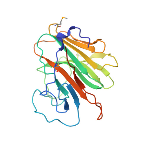
    2FBE

  • PubMed Abstract: 

    We determined the first structure of PRYSPRY, a domain found in over 500 different proteins, involved in innate immune signaling, cytokine signaling suppression, development, cell growth and retroviral restriction. The fold encompasses a 7-stranded and a 6-stranded antiparallel beta-sheet, arranged in a beta-sandwich. In the crystal, PRYSPRY forms a dimer where the C-terminus of an acceptor molecule binds to the concave surface of a donor molecule, which represents a putative interaction site. Mutations in the PRYSPRY domains of Pyrin, which are responsible for familial Mediterranean fever, map on the putative PRYSPRY interaction site.


  • Organizational Affiliation

    Department of Biochemistry, University of Zürich, CH-8057 Zürich, Switzerland.


Macromolecules
Find similar proteins by:  (by identity cutoff)  |  3D Structure
Entity ID: 1
MoleculeChains Sequence LengthOrganismDetailsImage
PREDICTED: similar to ret finger protein-like 1
A, B, C, D
201Homo sapiensMutation(s): 3 
Gene Names: gene 19q13.4.1
UniProt & NIH Common Fund Data Resources
Find proteins for A6NLU0 (Homo sapiens)
Explore A6NLU0 
Go to UniProtKB:  A6NLU0
PHAROS:  A6NLU0
GTEx:  ENSG00000223638 
Entity Groups  
Sequence Clusters30% Identity50% Identity70% Identity90% Identity95% Identity100% Identity
UniProt GroupA6NLU0
Sequence Annotations
Expand
  • Reference Sequence
Small Molecules
Modified Residues  1 Unique
IDChains TypeFormula2D DiagramParent
MSE
Query on MSE
A, B, C, D
L-PEPTIDE LINKINGC5 H11 N O2 SeMET
Experimental Data & Validation

Experimental Data

  • Method: X-RAY DIFFRACTION
  • Resolution: 2.52 Å
  • R-Value Free: 0.277 
  • R-Value Work: 0.227 
  • R-Value Observed: 0.227 
  • Space Group: P 41 2 2
Unit Cell:
Length ( Å )Angle ( ˚ )
a = 77.22α = 90
b = 77.22β = 90
c = 297.299γ = 90
Software Package:
Software NamePurpose
CNSrefinement
HKL-2000data reduction
SCALEPACKdata scaling
SHARPphasing

Structure Validation

View Full Validation Report


Entry History 

Deposition Data

Revision History  (Full details and data files)

  • Version 1.0: 2006-01-10
    Type: Initial release
  • Version 1.1: 2008-05-01
    Changes: Version format compliance
  • Version 1.2: 2011-07-13
    Changes: Version format compliance