2DCY

Crystal structure of Bacillus subtilis family-11 xylanase


Experimental Data Snapshot

  • Method: X-RAY DIFFRACTION
  • Resolution: 1.40 Å
  • R-Value Free: 0.217 
  • R-Value Work: 0.197 
  • R-Value Observed: 0.198 

wwPDB Validation   3D Report Full Report


This is version 2.1 of the entry. See complete history


Literature

Thermal Stabilization of Bacillus subtilis Family-11 Xylanase by Directed Evolution

Miyazaki, K.Takenouchi, M.Kondo, H.Noro, N.Suzuki, M.Tsuda, S.

(2006) J Biol Chem 281: 10236-10242

  • DOI: https://doi.org/10.1074/jbc.M511948200
  • Primary Citation of Related Structures:  
    2DCY, 2DCZ

  • PubMed Abstract: 

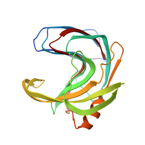
    We used directed evolution to enhance the thermostability of glycosyl hydrolase family-11 xylanase from Bacillus subtilis. By combining random point mutagenesis, saturation mutagenesis, and DNA shuffling, a thermostable variant, Xyl(st), was identified which contained three amino acid substitutions: Q7H, N8F, and S179C. The half-inactivation temperature (the midpoint of the melting curves) for the Xyl(st) variant compared with the wild-type enzyme after incubation for 10 min was elevated from 58 to 68 degrees C. At 60 degrees C the wild-type enzyme was inactivated within 5 min, but Xyl(st) retained full activity for at least 2 h. The stabilization was accompanied by evidence of thermophilicity; that is, an increase in the optimal reaction temperature from 55 to 65 degrees C and lower activity at low temperatures and higher activity at higher temperatures relative to wild type. To elucidate the mechanism of thermal stabilization, three-dimensional structures were determined for the wild-type and Xyl(st) enzymes. A cavity was identified around Gln-7/Asn-8 in wild type that was filled with bulky, hydrophobic residues in Xyl(st). This site was not identified by previous approaches, but directed evolution identified the region as a weak point. Formation of an intermolecular disulfide bridge via Cys-179 was observed between monomers in Xyl(st). However, the stability was essentially the same in the presence and absence of a reducing agent, indicating that the increased hydrophobicity around the Cys-179 accounted for the stability.


  • Organizational Affiliation

    Institute for Biological Resources and Functions, National Institute of Advanced Industrial Science and Technology (AIST), Central 6, 1-1-1 Higashi, Tsukuba, Ibaraki 305-8566, Japan. miyazaki-kentaro@aist.go.jp


Macromolecules
Find similar proteins by:  (by identity cutoff)  |  3D Structure
Entity ID: 1
MoleculeChains Sequence LengthOrganismDetailsImage
Endo-1,4-beta-xylanase A
A, B, C, D, E
185Bacillus subtilisMutation(s): 0 
Gene Names: xynA
EC: 3.2.1.8
UniProt
Find proteins for P18429 (Bacillus subtilis (strain 168))
Explore P18429 
Go to UniProtKB:  P18429
Entity Groups  
Sequence Clusters30% Identity50% Identity70% Identity90% Identity95% Identity100% Identity
UniProt GroupP18429
Sequence Annotations
Expand
  • Reference Sequence
Small Molecules
Ligands 3 Unique
IDChains Name / Formula / InChI Key2D Diagram3D Interactions
TAR
Query on TAR

Download Ideal Coordinates CCD File 
H [auth A]D(-)-TARTARIC ACID
C4 H6 O6
FEWJPZIEWOKRBE-LWMBPPNESA-N
TLA
Query on TLA

Download Ideal Coordinates CCD File 
G [auth A]
J [auth B]
K [auth B]
L [auth B]
N [auth C]
G [auth A],
J [auth B],
K [auth B],
L [auth B],
N [auth C],
O [auth E]
L(+)-TARTARIC ACID
C4 H6 O6
FEWJPZIEWOKRBE-JCYAYHJZSA-N
DIO
Query on DIO

Download Ideal Coordinates CCD File 
F [auth A],
I [auth B],
M [auth C]
1,4-DIETHYLENE DIOXIDE
C4 H8 O2
RYHBNJHYFVUHQT-UHFFFAOYSA-N
Experimental Data & Validation

Experimental Data

  • Method: X-RAY DIFFRACTION
  • Resolution: 1.40 Å
  • R-Value Free: 0.217 
  • R-Value Work: 0.197 
  • R-Value Observed: 0.198 
  • Space Group: P 21 21 2
Unit Cell:
Length ( Å )Angle ( ˚ )
a = 130.445α = 90
b = 186.209β = 90
c = 37.979γ = 90
Software Package:
Software NamePurpose
REFMACrefinement
HKL-2000data reduction
SCALEPACKdata scaling
MOLREPphasing

Structure Validation

View Full Validation Report


Entry History 

Deposition Data

Revision History  (Full details and data files)

  • Version 1.0: 2006-02-07
    Type: Initial release
  • Version 1.1: 2008-04-30
    Changes: Version format compliance
  • Version 1.2: 2011-07-13
    Changes: Version format compliance
  • Version 2.0: 2019-07-10
    Changes: Advisory, Atomic model, Data collection, Derived calculations, Non-polymer description, Structure summary
  • Version 2.1: 2023-10-25
    Changes: Data collection, Database references, Derived calculations, Refinement description