2CFM

ATP-DEPENDENT DNA LIGASE FROM PYROCOCCUS FURIOSUS


Experimental Data Snapshot

  • Method: X-RAY DIFFRACTION
  • Resolution: 1.80 Å
  • R-Value Free: 0.231 
  • R-Value Work: 0.204 
  • R-Value Observed: 0.204 

wwPDB Validation   3D Report Full Report


Ligand Structure Quality Assessment 


This is version 1.4 of the entry. See complete history


Literature

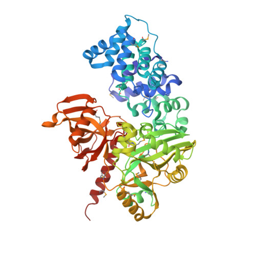
The Closed Structure of an Archaeal DNA Ligase from Pyrococcus Furiosus.

Nishida, H.Kiyonari, S.Ishino, Y.Morikawa, K.

(2006) J Mol Biol 360: 956

  • DOI: https://doi.org/10.1016/j.jmb.2006.05.062
  • Primary Citation of Related Structures:  
    2CFM

  • PubMed Abstract: 

    DNA ligases join single-strand breaks in double-stranded DNA, and are essential to maintain genome integrity in DNA metabolism. Here, we report the 1.8 A resolution structure of Pyrococcus furiosus DNA ligase (PfuLig), which represents the first full-length atomic view of an ATP-dependent eukaryotic-type DNA ligase. The enzyme comprises the N-terminal DNA-binding domain, the middle adenylation domain, and the C-terminal OB-fold domain. The architecture of each domain resembles those of human DNA ligase I, but the domain arrangements differ strikingly between the two enzymes. The closed conformation of the two "catalytic core" domains at the carboxyl terminus in PfuLig creates a small compartment, which holds a non-covalently bound AMP molecule. This domain rearrangement results from the "domain-connecting" role of the helical extension conserved at the C termini in archaeal and eukaryotic DNA ligases. The DNA substrate in the human open-ligase is replaced by motif VI in the Pfu closed-ligase. Both the shapes and electrostatic distributions are similar between motif VI and the DNA substrate, suggesting that motif VI in the closed state mimics the incoming substrate DNA. Two basic residues (R531 and K534) in motif VI reside within the active site pocket and interact with the phosphate group of the bound AMP. The crystallographic and functional analyses of mutant enzymes revealed that these two residues within the RxDK sequence play essential and complementary roles in ATP processing. This sequence is also conserved exclusively among the covalent nucleotidyltransferases, even including mRNA-capping enzymes with similar helical extensions at the C termini.


  • Organizational Affiliation

    Department of Structural Biology, Biomolecular Engineering Research Institute (BERI), Osaka 565-0874, Japan.


Macromolecules
Find similar proteins by:  (by identity cutoff)  |  3D Structure
Entity ID: 1
MoleculeChains Sequence LengthOrganismDetailsImage
THERMOSTABLE DNA LIGASE561Pyrococcus furiosusMutation(s): 0 
EC: 6.5.1.1
UniProt
Find proteins for P56709 (Pyrococcus furiosus (strain ATCC 43587 / DSM 3638 / JCM 8422 / Vc1))
Explore P56709 
Go to UniProtKB:  P56709
Entity Groups  
Sequence Clusters30% Identity50% Identity70% Identity90% Identity95% Identity100% Identity
UniProt GroupP56709
Sequence Annotations
Expand
  • Reference Sequence
Small Molecules
Ligands 2 Unique
IDChains Name / Formula / InChI Key2D Diagram3D Interactions
AMP
Query on AMP

Download Ideal Coordinates CCD File 
C [auth A]ADENOSINE MONOPHOSPHATE
C10 H14 N5 O7 P
UDMBCSSLTHHNCD-KQYNXXCUSA-N
MG
Query on MG

Download Ideal Coordinates CCD File 
B [auth A]MAGNESIUM ION
Mg
JLVVSXFLKOJNIY-UHFFFAOYSA-N
Modified Residues  1 Unique
IDChains TypeFormula2D DiagramParent
MSE
Query on MSE
A
L-PEPTIDE LINKINGC5 H11 N O2 SeMET
Experimental Data & Validation

Experimental Data

  • Method: X-RAY DIFFRACTION
  • Resolution: 1.80 Å
  • R-Value Free: 0.231 
  • R-Value Work: 0.204 
  • R-Value Observed: 0.204 
  • Space Group: P 1 21 1
Unit Cell:
Length ( Å )Angle ( ˚ )
a = 60.866α = 90
b = 87.399β = 109.22
c = 62.648γ = 90
Software Package:
Software NamePurpose
CNSrefinement
DENZOdata reduction
SCALEPACKdata scaling
SHARPphasing

Structure Validation

View Full Validation Report


Ligand Structure Quality Assessment 


Entry History 

Deposition Data

Revision History  (Full details and data files)

  • Version 1.0: 2006-07-12
    Type: Initial release
  • Version 1.1: 2011-05-08
    Changes: Version format compliance
  • Version 1.2: 2011-07-13
    Changes: Version format compliance
  • Version 1.3: 2019-01-30
    Changes: Data collection, Experimental preparation, Other
  • Version 1.4: 2019-02-06
    Changes: Data collection, Experimental preparation